
In-situ multiplex nucleic acid detection method

A technology of multiple nucleic acids and detection methods, applied in the field of molecular biology, can solve problems such as marker limitations and achieve good specificity

Active Publication Date: 2019-05-24
XIAMEN SEERNA BIOSCIENCE CO LTD
View PDF2 Cites 10 Cited by
  • Summary
  • Abstract
  • Description
  • Claims
  • Application Information

AI Technical Summary

Problems solved by technology

[0003] The above-mentioned traditional in situ hybridization nucleic acid detection method based on detection can only detect several different target nucleic acid sequences at the same time due to the limitation of the types of markers.

Method used


Image

  • In-situ multiplex nucleic acid detection method
  • In-situ multiplex nucleic acid detection method
  • In-situ multiplex nucleic acid detection method

Examples


Embodiment 1

[0026] Example 1 Human skin fibroblasts were used as experimental samples to investigate the in situ detection of cellular ACTB mRNA by double junction probe method.

[0027] (1) Cell culture and fixation:

[0028] The breast cancer cell line SK-BR-3 was cultured in DMEM (containing 10% FBS) for 24-48 hours, treated with trypsin to form suspended cells, planted on sterile glass slides, and cultured again for 12-24 hours. Rinse with DEPC-PBS, 3×3min; fix with 3% PFA prepared in DEPC-PBS at room temperature for 30min; rinse with DEPC-PBS again, and dehydrate with gradient ethanol: 70%, 85%, and 100%, respectively, for 5min each. Air dry.

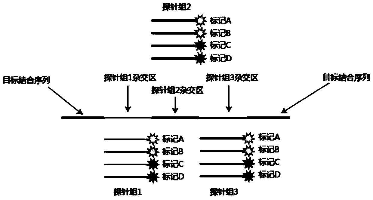
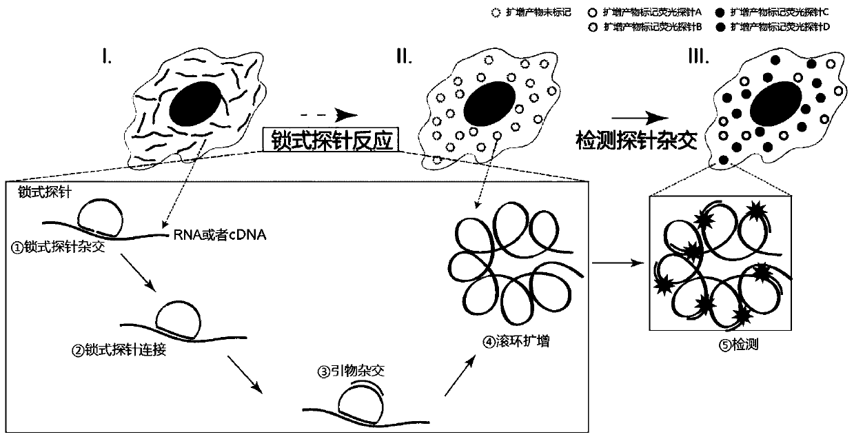
[0029] (2) In situ nucleic acid detection, such as Figure 1 to Figure 3 As shown, it specifically includes the following steps:

[0030] (1) padlock probe hybridization

[0031] The cell membrane of the breast cancer cell line SK-BR-3 was punched through the membrane, and 0.1M HCl was added to the sample and incubated at room temperature ...


PUM


Abstract

The invention discloses an in-situ multiplex nucleic acid detection method. The method highly integrates the fluorescence in situ hybridization technology with rolling circle amplification and probe coding technologies to realize highly multiplex in-situ detection of nucleic acid target sequences in samples. The in-situ multiplex nucleic acid detection method has the advantages that multiplex detection of target nucleic acids can be effectively realized, highly multiplex detection can be achieved, and the specificity is high.

Description

technical field [0001] The invention belongs to the technical field of molecular biology, and in particular relates to an in-situ multiple nucleic acid detection method. Background technique [0002] In situ hybridization technology is to realize heterohybridization between the labeled detection probe and the target nucleic acid sequence through complementary base pairing, the unbound detection probe is washed away, and then the detection probe bound to the target nucleic acid sequence is detected. In this way, the qualitative, localization and quantitative detection of the target nucleic acid can be realized in situ. In situ detection relies on the detection of a signal from the label of the detection probe. At present, the labels of detection probes mainly fall into two categories: fluorescent labels and biological enzyme labels. The former is generally a chemical molecule that can generate fluorescence, which is bound to the detection probe through covalent cross-linkin...

Claims


Application Information

Patent Timeline
Patent Type & Authority Applications(China)
IPC IPC(8): C12Q1/6841
Inventor 柯荣秦林辰陈小媛王自强刘玲姜萌
Owner XIAMEN SEERNA BIOSCIENCE CO LTD
Learn More