Quantitative method for determining sweetness at cellular level

A measurement method and quantitative measurement technology, applied in the field of molecular biology, can solve the problems of strong subjectivity in manual evaluation, inability to quantify sweetness, and inability to accurately quantify sweetness

Active Publication Date: 2019-01-04
CHINA TOBACCO YUNNAN IND
View PDF7 Cites 3 Cited by
  • Summary
  • Abstract
  • Description
  • Claims
  • Application Information

AI Technical Summary

Problems solved by technology

Analytical chemical methods can accurately detect the sugar content, but cannot quantify the sweetness at the physiological level; although biological detection methods can quantify the sweetness at the physiological level, manual evaluation is highly subjective and human tastes are different. difference, sweetness cannot be accurately quantified
At present, there is still no standard method for objectively quantifying the sweetness of substances at the physiological level. It is very important to establish a method that can quantify the sweetness of compounds at the physiological level for the development of food and sweeteners.

Method used


Image

  • Quantitative method for determining sweetness at cellular level
  • Quantitative method for determining sweetness at cellular level
  • Quantitative method for determining sweetness at cellular level

Examples


Embodiment 1

[0051] Changes of intracellular calcium ion concentration in HEK293 cells and HEK293-T1R2 / T1R3 cells under 150mM sucrose solution:

[0052] (1) Planting cells in a confocal culture dish:

[0053] Incubate the special confocal culture dish (outer diameter 35mm, inner diameter 10mm) with 1ml polylysine (25μg / ml) at 37°C for 30min, wash with phosphate buffer (10mM, pH 7.4) three times, and passage the cells in proportion into the confocal dish. After 24h-48h of adherent growth of the cells in the culture dish, it is observed under an optical microscope that the area of ​​the cells covering the bottom of the culture dish reaches 40%-60%.

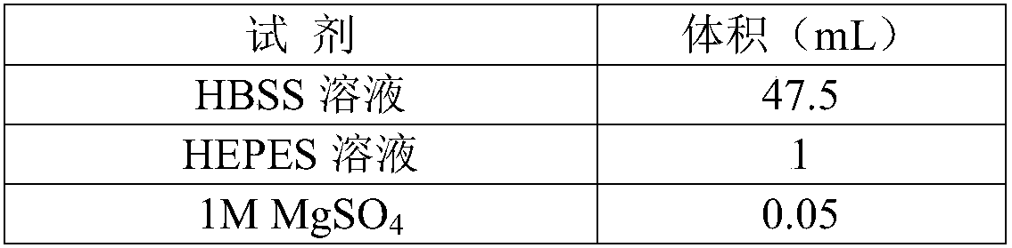
[0054] (2) Configure Flex buffer (pH=7.4)

[0055] Reagent

Volume (mL)

HBSS solution

47.5

HEPES solution

1

1M MgSO 4

0.05

1M Na 2 CO 3

0.165

1M CaCl 2

0.065

10% BSA bovine serum albumin

0.5

250mM Probenecid

0.5

total

50

[0056] The HBSS ...

Embodiment 2

[0076]Under the stimulation of 50 mM sucrose, the intracellular calcium ion concentration of HEK293 cells and HEK293-T1R2 / T1R3 cells changed, and the steps and conditions were the same as those in Example 1.

[0077] The fluorescence change ratio ΔR was obtained to be 0.105. If the sweetness of the 150 mM sucrose solution in Example 1 is set to 1, then the ΔR / ΔRs value is 0.370, and the relative sweetness of the 50 mM sucrose solution in this embodiment is 0.370.

Embodiment 3

[0079] Under the stimulation of 250 mM sucrose, the intracellular calcium ion concentration of HEK293 cells and HEK293-T1R2 / T1R3 cells changed, and the steps and conditions were the same as those in Example 1.

[0080] The fluorescence change ratio ΔR was obtained to be 0.487. Assuming that the sweetness of the 150mM sucrose solution in Example 1 is 1, the ΔR / ΔRs value is 1.71, and the relative sweetness of the 250mM sucrose solution in this example is 1.71.


PUM


Abstract

The invention provides a quantitative method for determining sweetness at a cellular level. The quantitative method for determining the sweetness at the cellular level includes various steps such as intracellular calcium ion fluorescence staining, laser confocal scanning, microscopic determination of intracellular calcium ion concentration changes. The quantitative method for determining the sweetness at the cellular level has the advantages of establishing a method for studying the intracellular calcium ion concentration changes of sweet receptor cells by using fluorescence detection technology, conducting quantitative perception of the sweetness of other unknown samples by humans in vitro, effectively avoiding the influence of human subjective factors, being fast, efficient, and highly sensitive in the detection of compound sweetness, and having a good application prospect.

Description

technical field [0001] The invention belongs to the field of molecular biology, and in particular relates to a method for quantitatively measuring sweetness at the cellular level. Background technique [0002] There are five main tastes that humans can distinguish, namely sour, sweet, bitter, umami, and salty. Most mammals prefer sweet tastes. Sweet foods usually indicate more energy, while bitter foods indicate that foods may be harmful to the body. Mammalian sweet taste receptor cells are mainly distributed on the taste bud cells of the tongue and palate epidermis. Taste signaling pathways are different in each type of cell that senses different tastes. [0003] The T1R gene family can encode three conserved GPCRs, namely T1R1, T1R2, and T1R3. T1R2 and T1R3 combine to form a heterodimer, which can act as a sweet taste receptor; T1R1 and T1R3 combine to form a heterodimer, which can serve as a fresh Taste receptors. These taste ligands interact with taste receptors, act...

Claims


Application Information

Patent Timeline
Patent Type & Authority Applications(China)
IPC IPC(8): G01N21/64
CPCG01N21/6428
Inventor 颜克亮苏莉翁俊陈微陈兴秦曦朱阳进尧晨光
Owner CHINA TOBACCO YUNNAN IND
Why Eureka
Try Eureka