
Plane splicing synthesis method of tissue slice microscopic images

A technology for microscopic images and tissue slices, applied in the field of image processing, to achieve the effect of overcoming the large number of image stitching, breaking through time-consuming and labor-intensive, and completing correctly

Inactive Publication Date: 2018-11-16
ANHUI NORMAL UNIV
View PDF1 Cites 12 Cited by
  • Summary
  • Abstract
  • Description
  • Claims
  • Application Information

AI Technical Summary

Problems solved by technology

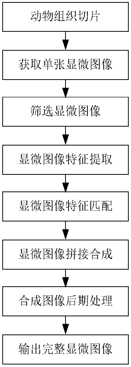
[0003] The purpose of the present invention is to overcome the deficiencies of the prior art and provide a method for plane splicing and synthesis of microscopic images of tissue slices to solve the deficiencies in the existing synthesis methods of microscopic images of slices and achieve efficient and accurate completion of microscopic images of animal tissue slices. Image plane splicing and synthesis

Method used


Image

  • Plane splicing synthesis method of tissue slice microscopic images
  • Plane splicing synthesis method of tissue slice microscopic images

Examples


Embodiment Construction

[0042] The specific implementation manner of the present invention will be described in further detail below by describing the best embodiment with reference to the accompanying drawings.

[0043] A method for plane splicing and synthesis of microscopic images of animal tissue sections, comprising the following steps:

[0044] (1) Acquisition of a single microscopic image. Tissue sections are made through the steps of tissue fixation, dehydration, transparency, paraffin embedding, sectioning, staining, and sealing, and then the prepared tissue sections are observed and photographed to obtain single microscopic images under different fields of view. Olympus BX51 optical microscope complete set of equipment was used to take microphotographs to obtain planar photos. Microscopic images were observed with the help of Image-Pro Plus 6.0 software on the computer, and several suitable and clear single-section microscopic images were selected for subsequent stitching and synthesis of ...


PUM


Abstract

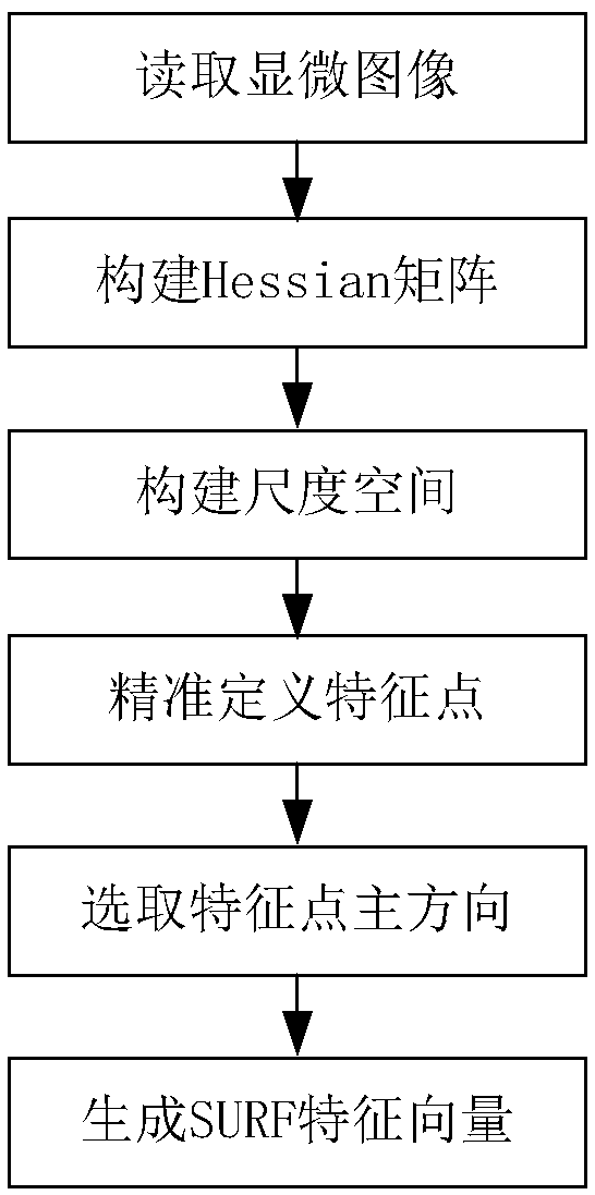
The invention discloses a plane splicing synthesis method of tissue slice microscopic images. The method comprises the following steps: (1) obtaining to-be-spliced microscopic images; (2) extracting features of the microscopic images; (3) matching the features of the microscopic images; (4) performing splicing synthesis on the microscopic images. The invention has the advantages of making up defects of a prior slice microscopic image synthesis method and achieving efficient and accurate plane splicing synthesis of animal tissue slice microscopic images.

Description

technical field [0001] The invention relates to the field of image processing, in particular to a method for plane splicing and synthesis of microscopic images of tissue sections. Background technique [0002] In the process of observation and 3D reconstruction of animal tissues, it is necessary to obtain complete microscopic images of tissue slices. However, in order to ensure that the images are clear and reach a certain pixel degree during the shooting process, it is impossible to directly obtain a complete tissue slice image by taking a single photo. It can take pictures of some areas of tissue slices under the microscope field of view. Therefore, in order to obtain a clear and complete microscopic image of a paraffin section, multiple high-resolution images need to be taken. In order to obtain a complete microscopic image, all photos need to be spliced ​​and synthesized to generate a complete high-resolution image. Microscopic images of tissue sections. At present, mo...

Claims


Application Information

Patent Timeline
Patent Type & Authority Applications(China)
IPC IPC(8): G06T3/40G06T5/50
CPCG06T3/4038G06T5/50G06T2207/10061
Inventor 刘再群李蓓陈梅芳
Owner ANHUI NORMAL UNIV
Learn More