Retina hemangioma image segmentation method based on GAN (Generative Adversarial Network)
A retinal blood vessel and image segmentation technology, applied in image analysis, biological neural network model, image enhancement, etc., can solve the problem of missing details of blood vessel branches, useless information, complex distribution of thick and thin blood vessels, and inability to take into account the integrity and accuracy of hemangioma. , to achieve the effect of reducing segmentation results, accurate segmentation details, and improving segmentation efficiency
- Summary
- Abstract
- Description
- Claims
- Application Information
AI Technical Summary
Problems solved by technology
Method used
Image
Examples
Embodiment 1
[0042] This embodiment discloses a retinal hemangioma segmentation method based on generating an adversarial network, such as figure 1 shown, including the following steps:
[0043] (1) Obtain training data
[0044] First, the fundus image of the retina is collected. The fundus image provides sufficient information and is widely used in disease diagnosis. The fundus image is directly obtained by the fundus camera. The fundus image is preprocessed, and the resolution of the image is unified to N×N, and the hemangioma in the image is manually and accurately segmented, and the segmented data is used for subsequent training.
[0045] (2) Construct Generative Adversarial Network
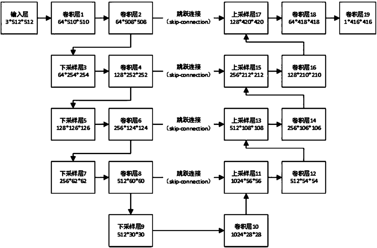
[0046] Construct generator G(x, the generator network includes at least a plurality of downsampling network layers, upsampling network layers with the same number of downsampling network layers. Wherein, the downsampling network layer includes a downsampling layer, a volume product layer and an activat...
Embodiment 2
[0060] The purpose of this embodiment is to provide a computing device.
[0061] In order to achieve the above object, the present invention adopts the following technical scheme:
[0062] A device for segmenting images of retinal hemangiomas based on generating an adversarial network, comprising a memory, a processor, and a computer program stored on the memory and operable on the processor, when the processor executes the program, it realizes:
[0063] Construct generation confrontation network structure, described generation confrontation network comprises generator and discriminator;
[0064] Using artificially segmented images of hemangioma and original fundus retinal images as training data, iteratively trains the generated confrontation network to obtain an optimal generator;
[0065] The hemangioma image is segmented based on the optimal generator for the fundus retinal image to be segmented.
Embodiment 3
[0067] The purpose of this embodiment is to provide a computer-readable storage medium.
[0068] In order to achieve the above object, the present invention adopts the following technical scheme:
[0069] A computer-readable storage medium having stored thereon a computer program which, when executed by a processor, performs:
[0070] Construct generation confrontation network structure, described generation confrontation network comprises generator and discriminator;
[0071] Using artificially segmented images of hemangioma and original fundus retinal images as training data, iteratively trains the generated confrontation network to obtain an optimal generator;
[0072] The hemangioma image is segmented based on the optimal generator for the fundus retinal image to be segmented.
PUM
 Abstract
Description
Claims
Application Information
 - Generate Ideas
- Intellectual Property
- Life Sciences
- Materials
- Tech Scout
- Unparalleled Data Quality
- Higher Quality Content
- 60% Fewer Hallucinations
Browse by: Latest US Patents, China's latest patents, Technical Efficacy Thesaurus, Application Domain, Technology Topic, Popular Technical Reports.
© 2025 PatSnap. All rights reserved.Legal|Privacy policy|Modern Slavery Act Transparency Statement|Sitemap|About US| Contact US: help@patsnap.com


