Device special for radiology department

A technology for radiology and equipment, applied in the field of medical equipment, can solve the problems of confusion, fading, and increase the difficulty of work for medical personnel, and achieve the effect of convenient management and ease of work difficulty.

Inactive Publication Date: 2018-05-25
十堰利兹传媒有限公司
View PDF0 Cites 0 Cited by
  • Summary
  • Abstract
  • Description
  • Claims
  • Application Information

AI Technical Summary

Problems solved by technology

[0002] Traditional medical imaging image output uses paper or other media, which has no relevant characteristics such as wear resistance, pressure resistance, and translucency. Due to its soft texture, it is easy to curl during handheld observation, and the quality of image output is not very good. After a period of time under the irradiation of light, the image will turn yellow, fade, fade, and the image will be blurred, which is very troublesome to operate and not conducive to scientific medical diagnosis
[0003] Most of the diagnostic reports are separated from the imaging film when performing radiographic diagnosis for patients, so it is easy to lose or confuse the report. Personnel increase the difficulty of work

Method used


Image

  • Device special for radiology department
  • Device special for radiology department
  • Device special for radiology department

Examples


Embodiment

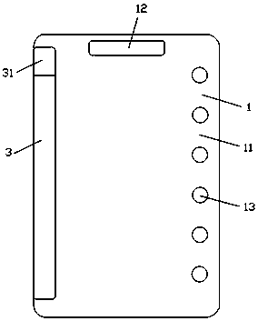
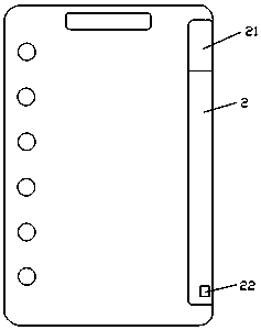
[0018] The embodiment of the present invention provides a kind of radiology special equipment, see figure 1 , figure 2 , the special radiology equipment includes an image film 1, a reinforcement sheet 2 arranged at one end of the image film 1, and a magnetic sheet 3 arranged at the other end of the image film 1;

[0019] The video film 1 includes a film body 11, a handle hole 12 arranged at the end of the film body 11 in sequence, and a placement hole 13 arranged at the side of the film body 11; the handle hole 12 is provided to facilitate people to carry; the placement hole 13 is arranged to facilitate overall storage and carrying ;

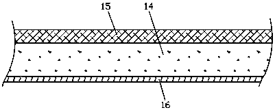
[0020] see image 3 The film body 11 includes a paper substrate 14, a protective layer 15 coated on one side of the paper substrate 14, and a plastic-coated layer 16 coated on the other side of the paper substrate 14;

[0021] The protective layer 15 includes 85% self-duplex resin and 15% water-based acrylic emulsion; making the film antist...


PUM


Abstract

The invention relates to a device special for the radiology department, and belongs to the field of medical devices. The device special for the radiology department comprises image film, a reinforcement sheet arranged at the end of one side of the image film and a magnetic sheet arranged at the end of the other side of the image film. According to the device, an adhesion phenomenon cannot be generated between the film and the film; a handle hole and placing holes are formed and are beneficial to integral storing and carrying, and the reinforcement sheet is arranged and can prevent curling of the image film; a label containing groove and a U disk containing groove are formed and can contain basis information and electronic image data of a patient; and meanwhile, the magnetic sheet and the reinforcement sheet tightly attract each other and can clamp other medical treatment data of the patient together with the film, the medical treatment data of the patient can be subjected to unified classification management with the patient information in time, convenience is brought to management on the medical treatment data of the patient, and the working difficulty of medical personnel of theradiology department is reduced.

Description

technical field [0001] The invention relates to the field of medical equipment, in particular to a special radiology equipment. Background technique [0002] Traditional medical imaging image output uses paper or other media, which has no relevant characteristics such as wear resistance, pressure resistance, and translucency. Due to its soft texture, it is easy to curl during handheld observation, and the quality of image output is not very good. After a period of time under the irradiation of light, the image will turn yellow, lose color, fade, and blur the image, which is very troublesome to operate and not conducive to scientific medical diagnosis. [0003] Most of the diagnostic reports are separated from the imaging film when performing radiographic diagnosis for patients, so it is easy to lose or confuse the report. Personnel increase the difficulty of work. Contents of the invention [0004] In order to solve the problems of the prior art, the embodiment of th...

Claims


Application Information

Patent Timeline
Patent Type & Authority Applications(China)
IPC IPC(8): G03B42/04
CPCG03B42/047
Inventor 徐韬
Owner 十堰利兹传媒有限公司
Why Eureka
Try Eureka