
Acoustic microscopic imaging device for tooth body

A technology of microscopic imaging and tooth body, which is applied in the direction of measuring device, analysis of solids with sound wave/ultrasonic wave/infrasonic wave, material analysis with sound wave/ultrasonic wave/infrasonic wave, etc. Observation effect, complex sample preparation process, time-consuming and other problems, to achieve the effect of rapid diagnosis and inspection and environmental protection

Active Publication Date: 2017-12-26
AVIC BASIC TECH RES INST
View PDF7 Cites 1 Cited by
  • Summary
  • Abstract
  • Description
  • Claims
  • Application Information

AI Technical Summary

Problems solved by technology

Its main disadvantages are: 1) Visual inspection tools can not get the internal information of dental tissue, especially it is difficult to grasp the internal changes or pathological information of some early dental tissue, which may lose the best dental treatment Timing; 2) Micro-X-ray CT equipment has obvious radiation damage to patients, and it is difficult to obtain information on internal changes or lesions of early dental tissues
Its main disadvantages are: 1) It can only observe the tissue with optical gray scale difference on the surface of the sample and its changes; 2) It cannot obtain information on the internal changes or lesions of the internal tooth tissue and early tooth tissue of the tooth sample; 3 ) The mechanical sectioning process will cause damage to the tooth sample, which will affect the actual observation effect, and it is difficult to distinguish whether the microscopic damage in the tooth body originally existed or was generated during the mechanical sectioning process; 4) Complex sample preparation is required process, time-consuming

Method used


Image

  • Acoustic microscopic imaging device for tooth body
  • Acoustic microscopic imaging device for tooth body

Examples


Embodiment

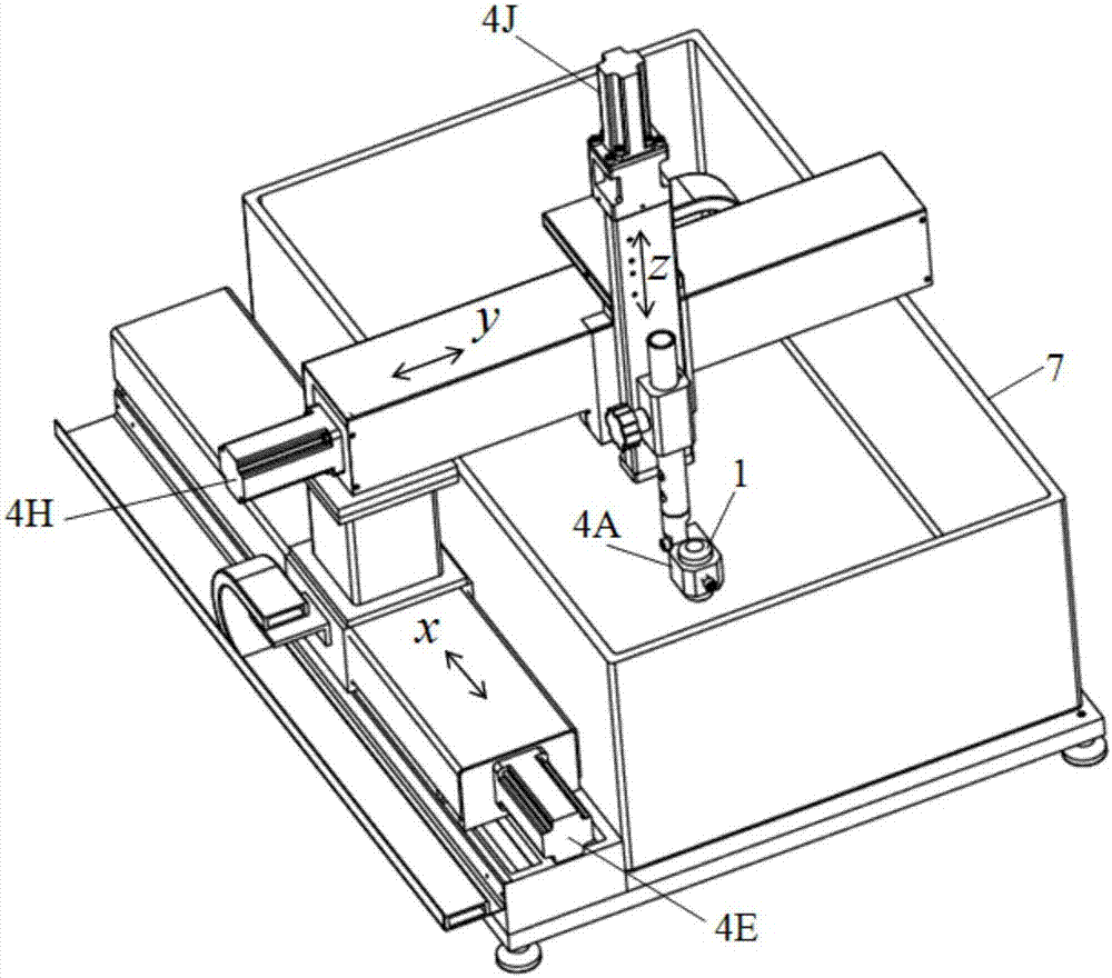
[0051] The x, y, z, A four-axis scanning mechanism of AVIC Composite Materials Co., Ltd. is selected, the micro-stepping motor and its control unit are used as the acoustic scanning control unit, and the high-frequency ultrasonic unit of AVIC Composite Materials Co., Ltd. is selected as the acoustic excitation / reception Unit, select the acoustic lens of 50MHz and 100MHz, adopt the data acquisition system of 1GMz and 2GHz as the acoustic information processing unit, adopt the computer of 1.5GHz main frequency, 512M internal memory as the host computer of the acoustic imaging unit, construct dental body acoustic microscope according to the present invention The imaging device uses acoustic lenses with focal points of 25 microns and 50 microns, and adopts plane scanning and cross-sectional scanning to analyze and apply a series of actual acoustic microscopic imaging experiments on actual human tooth samples from different clinics. The application results show that using The constr...


PUM


Abstract

The invention belongs to the technical field of medical science and nondestructive testing, and relates to an acoustic microscopic imaging device for a tooth body. The acoustic microscopic imaging device for the tooth body comprises an acoustic lens, an acoustic excitation / receiving unit, an acoustic information processing unit, an acoustic scanning mechanism, an acoustic scanning control unit, an acoustic imaging unit and a fixed platform. According to the acoustic microscopic imaging device, the acoustic scanning mechanism is utilized to enable the acoustic lens to perform acoustic micro scanning on the tooth body and extract an acoustic wave signal and a scanning position signal of the inner part of the detected tooth body in real time; the signals are used for 2D (two dimensional) and 3D (three dimensional) acoustic microscopic imaging display of the tooth body, so that acoustic microscopic imaging of the surface and internal tissues of the tooth body is realized, and an acoustic microscopic scanning resolution ratio can reach 10 microns. Experimental application results show that the acoustic microscopic imaging device disclosed by the invention can rapidly obtain the characteristic distribution of tissues of the tooth body such as internal enamel, dentin, dental pulp and cementum and changes of the tissues of the tooth body, and can obtain relatively good actual acoustic imaging effects.

Description

technical field [0001] The invention belongs to the technical field of medicine and non-destructive testing, and relates to an acoustic microscopic imaging device for teeth. Background technique [0002] Changes in dental tissue are the main root cause of dental cavity diseases. Usually, when there are obvious lesions in dental tissue, it may cause tooth function failure. At present, clinically, the attending doctor mainly uses visual tools or auxiliary Micro-X-ray CT equipment to check and diagnose tooth damage or tooth disease. Its main disadvantages are: 1) Visual inspection tools can not get the internal information of dental tissue, especially it is difficult to grasp the internal changes or pathological information of some early dental tissue, which may lose the best dental treatment Timing; 2) Micro-X-ray CT equipment has obvious radiation damage to patients, and it is also difficult to obtain information on internal changes or lesions of early dental tissues. In or...

Claims


Application Information

Patent Timeline
IPC IPC(8): G01N29/06G01N29/04
CPCG01N29/045G01N29/0681G01N2291/023G01N2291/0289
Inventor 刘松平刘菲菲李乐刚白金鹏
Owner AVIC BASIC TECH RES INST
Learn More