
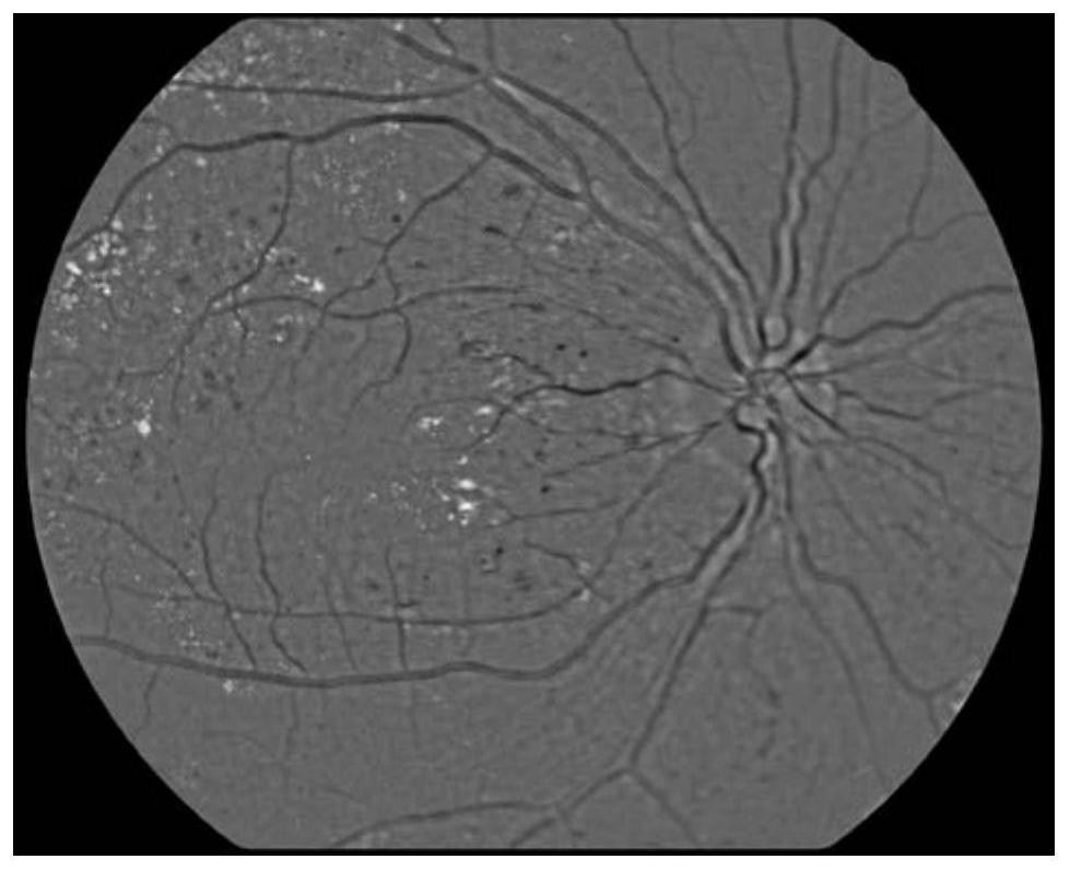
A method for quality control of fundus images

A quality control method and fundus image technology, applied in the field of image processing, can solve problems such as troublesome detection and repetitive work, and achieve the effect of reducing technical requirements for shooting and saving feedback time

Active Publication Date: 2020-11-06
北京大恒普信医疗技术有限公司
View PDF10 Cites 0 Cited by
  • Summary
  • Abstract
  • Description
  • Claims
  • Application Information

AI Technical Summary

Problems solved by technology

[0005] Therefore, the existing image acquisition equipment will bring a lot of unnecessary trouble and repetitive work to the follow-up detection

Method used


Image

  • A method for quality control of fundus images
  • A method for quality control of fundus images
  • A method for quality control of fundus images

Examples


Embodiment Construction

[0046] The present invention will be described in detail below with reference to the accompanying drawings and the embodiments thereof, but the protection scope of the present invention is not limited to the scope described in the embodiments.

[0047] Such as figure 1 Shown is a flowchart of an image quality control method according to an embodiment of the present invention. It should be noted that the flow chart of this embodiment is the most detailed quality control process, but those skilled in the art should understand that in some application scenarios, such comprehensive quality control is not required, and these steps can be combined arbitrarily to form Simplified quality control methods.

[0048] Firstly, the RGB image data of the target fundus image is obtained, and then the following image quality judgment, screening and processing are performed.

[0049] Single-channel image discrimination: In order to obtain a color fundus image with rich information, the method...


PUM


Abstract

The invention discloses an eye fundus image quality control method which includes the following steps: processing a target eye fundus image; extracting local bright regions and local dark regions of the target eye fundus image from a region of interest ROI of the target eye fundus image; determining whether the brightness of the target eye fundus image is abnormal; and carrying out image screening based on the judgment on brightness. Through the method, the quality of color eye fundus images can be discriminated, and images with local bright regions and / or local dark regions can be distinguished from normal eye fundus images. The quality of color eye fundus images taken by cameras of various models can be controlled, evaluated and classified. The visual area of an eye fundus image can be identified, and the quality of the eye fundus image can be evaluated. Unqualified images are prevented from entering the detection link to occupy resources. The method is used together with eye fundus camera hardware. Problems can be found at the source, and the feedback time can be reduced. The shooting technology requirement for medical workers is lowered.

Description

technical field [0001] The invention relates to the field of image processing, in particular to a method for quality screening of color fundus images. Background technique [0002] In the detection process of eye lesions, color fundus images are a very important detection basis. [0003] In the screening of color fundus image lesions, doctors attach great importance to the visual area of ​​the fundus image. Only a meaningful visual area can reflect the presence or absence of lesions and the degree of lesions. For example, the detection of myopia should focus on the vicinity of the optic disc, whether there are changes in the shape and size of the optic disc and the optic cup, and to what extent. The detection of macular edema should focus on the macular area. According to the diagnostic experience of first-line clinical ophthalmologists, when it is required to collect images, each patient's left and right eyes must have one image centered on the optic disc and one image c...

Claims


Application Information

Patent Timeline
Patent Type & Authority Patents(China)
IPC IPC(8): G06T7/00
CPCG06T7/0002G06T7/0012G06T2207/10024G06T2207/30041G06T2207/30168
Inventor 赵雷唐轶
Owner 北京大恒普信医疗技术有限公司
Learn More