Head-mounted slit lamp micrographic examination system

An inspection system and slit lamp technology, applied in the fields of medical science, ophthalmoscope, eye testing equipment, etc., can solve the problems of difficult semi-self-service or self-help inspection, inconvenience of carrying, operation, inconvenience of inspection and health care, etc., to achieve easy operation , The effect of facilitating daily routine inspection and simplifying equipment composition

Inactive Publication Date: 2017-08-04
XIAMEN UNIV
View PDF10 Cites 3 Cited by
  • Summary
  • Abstract
  • Description
  • Claims
  • Application Information

AI Technical Summary

Problems solved by technology

[0004] To sum up, the slit lamps currently on the market are either poor in real-time performance and difficult to quickly detect; or they are inconvenient to carry and operate, and it is difficult to realize semi-self-service or self-service inspection without on-site doctors, and it is not convenient for daily routine inspection and health care

Method used


Image

  • Head-mounted slit lamp micrographic examination system

Examples


Embodiment Construction

[0013] The specific embodiments of the present invention will be described in detail below in conjunction with the accompanying drawings.

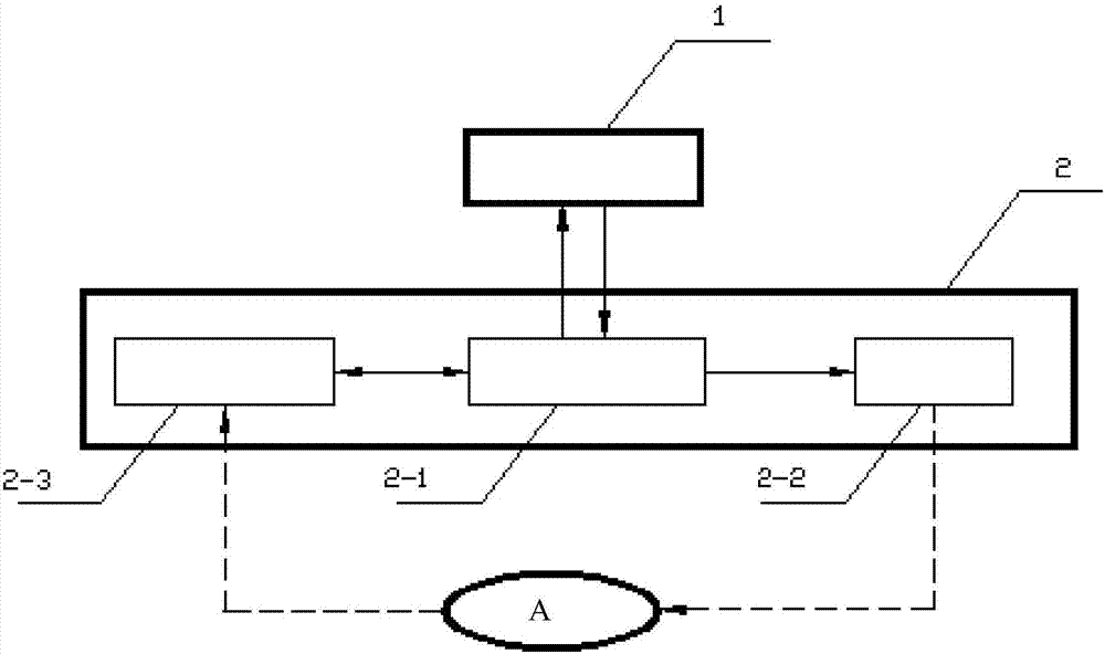
[0014] Such as figure 1 As shown, the embodiment of the present invention is provided with an inspection operation terminal 1 and a head-mounted unit 2 . The medical personnel set the detection control information (including information such as the width, color, and shape of the detection spot) and image acquisition information (magnification, etc.) required for the slit lamp microscope detection at the inspection operation terminal 1 . After the information is processed to form a patterned control command, it is sent to the head-mounted unit 2 through the communication network. After the head-mounted unit 2 receives the control command, the central control module 2-1 analyzes and processes the command to form a light source control command and an image acquisition command. , and send the light source control instruction to the projection...


PUM


Abstract

A head-mounted slit lamp microscopy system relates to the field of slit lamp microscopy. There is an inspection operation terminal and a head-mounted unit; the head-mounted unit is equipped with a projection module, an image acquisition module and a central control module, and the projection module is equipped with a projection drive part and a lighting part; Check the light band information to form a patterned control command, and send the patterned control command to the head-mounted unit; the central control module analyzes and processes the patterned command, sends the light source control command to the projection module, and the image acquisition command to the image acquisition module; the projection module According to the light source control instruction, the detection spot is generated and projected to the patient's eye; the image acquisition module collects the optical slice image of the inspection part according to the image acquisition instruction, and sends the optical slice image data to the central control module; the central control module according to the received The optical slice image data forms an inspection data packet, and the inspection data packet is sent to the inspection operation end.

Description

technical field [0001] The invention relates to the field of slit lamp microscopy, in particular to a head-mounted slit lamp microscopic inspection system for eye disease inspection. Background technique [0002] The slit lamp microscope is composed of a binocular microscope and an illumination system. Its working principle is: irradiate the high-brightness slit light band to the inspected part of the eye at a suitable angle, and obtain an optical section of the living tissue for observation to diagnose whether the eyeball has occurred. It is an important instrument commonly used in the examination and diagnosis of ophthalmic diseases. [0003] The traditional slit lamp consists of an illumination system, a binocular stereo microscope (observation system), a moving slide system and a workbench (base). From the structure, there are desktop, portable and so on. These structures require constant adjustment of the slit lamp during use to obtain optical sections at correspondin...

Claims


Application Information

Patent Timeline
IPC IPC(8): A61B3/135A61B3/14
CPCA61B3/135A61B3/0008A61B3/0083A61B3/14
Inventor 陈延平肖啸杨英迪颜黄苹朱相宇
Owner XIAMEN UNIV
Why Eureka
Try Eureka