
Calprotectin participating in in-vitro binding of NY-ESO-1 and DC cells in bone marrow

A technology of NY-ESO-1 and calreticulin, applied in the field of biomedicine, can solve problems such as affecting the natural history of tumors

Inactive Publication Date: 2017-05-31
宁波美丽人生医药生物科技发展有限公司
View PDF0 Cites 0 Cited by
  • Summary
  • Abstract
  • Description
  • Claims
  • Application Information

AI Technical Summary

Problems solved by technology

Furthermore, the reduction of spontaneous NY-ESO-1 antigen in melanoma suggests that immune responses against NY-ESO-1 may influence the natural history of tumors

Method used


Image

  • Calprotectin participating in in-vitro binding of NY-ESO-1 and DC cells in bone marrow
  • Calprotectin participating in in-vitro binding of NY-ESO-1 and DC cells in bone marrow
  • Calprotectin participating in in-vitro binding of NY-ESO-1 and DC cells in bone marrow

Examples


Embodiment 1

[0065] NY-ESO-1 protein can form multimeric structures even in loading buffer in the presence of regular concentrations of β-mercaptoethanol.

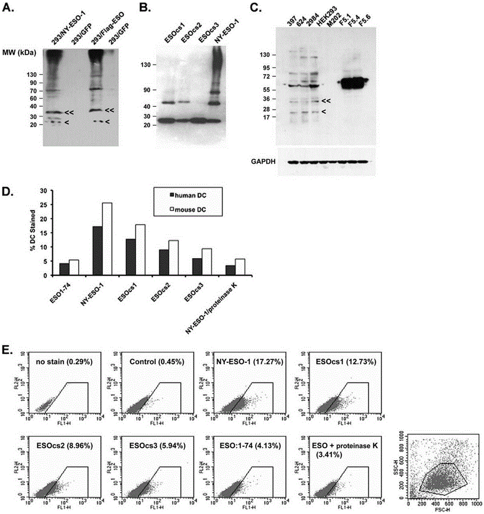
[0066] Experiments have found that NY-ESO-1 proteins obtained from bacteria and mammalian cells can form multimeric structures even in loading buffers with conventional concentrations of β-mercaptoethanol ( figure 1 A, B).

Embodiment 2

[0068] The polymerization reaction of NY-ESO-1 is mediated by its intermolecular disulfide bonds.

[0069] Cysteine ​​in amino acid residues 75, 76, 78, 152 and 165 were replaced with serine. Three types of NY-ESO-1 with amino acid substitutions were: ESOcs1 with cysteine-serine substitutions at positions 75, 76, and 78, ESOcs2 with substitutions at positions 152 and 165, and ESOcs3 with mutations at all five positions. Purify these proteins and use Western blot to compare the oligomerization state between them ( figure 1 B). The results showed that the wild-type protein clearly showed the presence of dimers, tetramers and even multimers of 130 kDa. ESOcs1 and ESOcs2 showed mainly monomer and dimer structures, while the result of ESOcs3 showed mainly monomer. This experiment shows that the polymerization of NY-ESO-1 is due to the disulfide bonds between its molecules.

Embodiment 3

[0071] NY-ESO-1 binds to human and mouse immature dendritic cells in vitro and is related to its own polymeric structure.

[0072] Purify these proteins and use Western blot to compare the oligomerization state between them ( figure 1 B). The results showed that the wild-type protein clearly showed the presence of dimers, tetramers and even multimers of 130 kDa. ESOcs1 and ESOcs2 showed mainly monomer and dimer structures, while the result of ESOcs3 showed mainly monomer. This experiment shows that the polymerization of NY-ESO-1 is due to the disulfide bonds between its molecules. NY-ESO-1 present in cancer cells was also analyzed by Western blot for its protein aggregation in the loading buffer containing conventional concentrations of β-mercaptoethanol, and it was found that NY-ESO-1 mainly manifested as trimers and tetramers Dimeric and monomeric forms are rarely found (mutation 1C). Experiments show that NY-ESO-1 binds to human and mouse immature dendritic cells in vit...


PUM

PropertyMeasurementUnit
molecular weightaaaaaaaaaa

Abstract

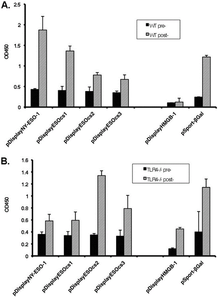
The invention relates to the field of biological medicine, in particular to calprotectin participating in in-vitro binding of NY-ESO-1 and DC cells in bone marrow. The inventor of the invention explores that the NY-ESO-1 protein can form a polymer structure even in loading buffer with beta-mercaptoethanol with the conventional concentration; the polymerization reaction of the NY-ESO-1 is mediated by intermolecular disulfide bond; binding of the NY-ESO-1 and immature dendritic cells of human and mice in vitro is associated with the polymerization structure of the NY-ESO-1; TLR4 influences the in-vitro binding of the NY-ESO-1 and the DC cells in the bone marrow; the binding of the NY-ESO-1 and the immature dendritic cells of human and the mice in vitro can be realized through the effect of the calprotectin; polymer oligomerization reduces the binding quantity of the TLR4 and the NY-ESO-1; the polymer structure of the NY-ESO-1 and the TLR4 in a host participate in the immuno-sphere antibody reaction; and improvement of Artv1(wtArt) and CA9 gene immunogenicity depends on the NY-ESO-1 gene fusion expression. The invention proves that the calprotectin participates in in-vitro binding of the NY-ESO-1 and the DC cells in the bone marrow.

Description

technical field [0001] The invention relates to the field of biomedicine, in particular to calreticulin participating in the combination of NY-ESO-1 and DC cells in bone marrow in vitro. Background technique [0002] It has been postulated that innate immune system receptors may be involved in the initial autoimmune response to tumor-specific antigens (TAAs). Necrotic tumor cells release endogenous factors or damage-associated pattern molecules such as heat shock proteins, high mobility group box B1 (HMGB-1), and uric acid. These damage-patterning molecules alert the innate immune system via CD91, receptor for advanced glycation end products, TLR2 / TLR4, and interleukin-1 (IL-1) receptor, respectively. TAA itself is generally regarded as a bystander of the above-mentioned "danger signal", acting as an adjuvant in the initial stage of the anti-tumor immune response. According to this example, the spontaneous immune response is caused by new peptides produced by mutations in ...

Claims


Application Information

Patent Timeline
Patent Type & Authority Applications(China)
IPC IPC(8): C12N5/0784C07K14/47
Inventor 田晓丽
Owner 宁波美丽人生医药生物科技发展有限公司
Learn More