Method for processing cardiac perfusion magnetic resonance image

A magnetic resonance image and processing method technology, applied in the field of medical images, can solve the problems of complicated semi-automatic detection methods, low efficiency, and large inter-observer, and achieve the effect of visualization and fully automatic quantitative analysis

Active Publication Date: 2017-05-24
SHANGHAI UNITED IMAGING HEALTHCARE
View PDF3 Cites 8 Cited by
  • Summary
  • Abstract
  • Description
  • Claims
  • Application Information

AI Technical Summary

Problems solved by technology

Among them, manual and user-assisted semi-automatic detection methods are more complicated, inefficient, and there are large differences between observers and observers

Method used


Image

  • Method for processing cardiac perfusion magnetic resonance image
  • Method for processing cardiac perfusion magnetic resonance image
  • Method for processing cardiac perfusion magnetic resonance image

Examples


Embodiment Construction

[0039] The present invention will be further described below in conjunction with the accompanying drawings and embodiments.

[0040] A method for processing cardiac perfusion magnetic resonance images, comprising the following main steps:

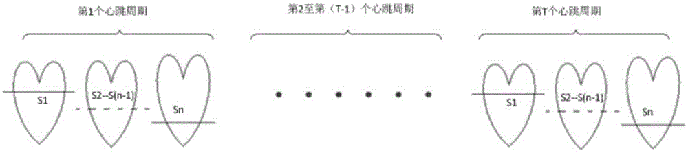
[0041] Acquisition of an MRI image containing several slices of the left ventricular myocardium I NT , where N represents the sequence number of the slice layer in the same heartbeat cycle, T represents the sequence number of different heartbeat cycles, and both N and T are integers greater than or equal to 1;

[0042] Starting from the initial layer, the magnetic resonance image of N slices is divided into the endocardium within T heartbeat cycles, and the corresponding magnetic resonance image I of each slice is obtained. MT The reference image I Mr , where M is an integer greater than or equal to 1 and less than or equal to N;

[0043] MR image in slices per slice I Mr The reference image in I Mr As a benchmark, complete the registr...


PUM


Abstract

The invention discloses a method for processing a cardiac perfusion magnetic resonance image. The method comprises the steps of: acquiring magnetic resonance images INT of a plurality of layers of slices including left ventricular myocardium; from the start layer, respectively segmenting the myocardial inner membrane of the magnetic resonance images of the N layers of slices within T heartbeat cycles to obtain a reference image Imr corresponding to each layer of slice; based on the reference image Imr, registering the magnetic resonance image of the current layer of slice within the T heartbeat cycles; based on the segmentation result of the myocardial inner membrane of the magnetic resonance image of each layer of slice, segmenting a myocardial outer membrane; calculating and smoothing a mean gray-time curve of a left ventricular cavity zone and a right ventricular cavity zone; and taking the connecting line of the centroid point of a blood pool zone defined by the myocardial inner membrane and the center point of a right ventricular boundary as a base line, blocking the myocardium according to a bovine eye graph partitioning method. The method can realize visual and full-automatic quantitative analysis on the signal intensity change curve of the left ventricular myocardium with time.

Description

[0001] 【Technical field】 [0002] The invention relates to the field of medical images, in particular to a post-processing method for cardiac perfusion magnetic resonance images. [0003] 【Background technique】 [0004] In recent years, the morbidity and mortality of cardiovascular diseases are increasing year by year. Tens of millions of people die of cardiovascular disease every year all over the world. Myocardial ischemia is the most important cause of cardiovascular morbidity and mortality. Cardiac perfusion magnetic resonance imaging is considered the first-line noninvasive test for ischemic heart disease. According to the basic principles of cardiac perfusion magnetic resonance images, radiologists can make a clinical diagnosis of myocardial ischemia by directly observing the changes in the signal intensity of the myocardial part of the left ventricle over time. However, deformation and motion artifacts in cardiac perfusion MR images caused by cardiac motion, respirati...

Claims


Application Information

Patent Timeline
Patent Type & Authority Applications(China)
IPC IPC(8): G06T7/11
CPCG06T2207/10088G06T2207/20104G06T2207/30048
Inventor 姜娈凌姗李强
Owner SHANGHAI UNITED IMAGING HEALTHCARE
Why Eureka
Try Eureka