
Aneurysm model based on 3D printing and manufacturing method thereof

A 3D printing and aneurysm technology is applied in the field of preparing aneurysm models for teaching and surgical training, which can solve the problems of complex process and many steps, and achieve the effect of simple process.

Inactive Publication Date: 2017-05-17
李翔宇
View PDF4 Cites 14 Cited by
  • Summary
  • Abstract
  • Description
  • Claims
  • Application Information

AI Technical Summary

Problems solved by technology

This processing method has many steps and complicated process. It is used to mass-produce standardized normal human body structure. It is not suitable for individualized, complicated and diversified disease models.

Method used


Image

  • Aneurysm model based on 3D printing and manufacturing method thereof
  • Aneurysm model based on 3D printing and manufacturing method thereof
  • Aneurysm model based on 3D printing and manufacturing method thereof

Examples


Embodiment 1

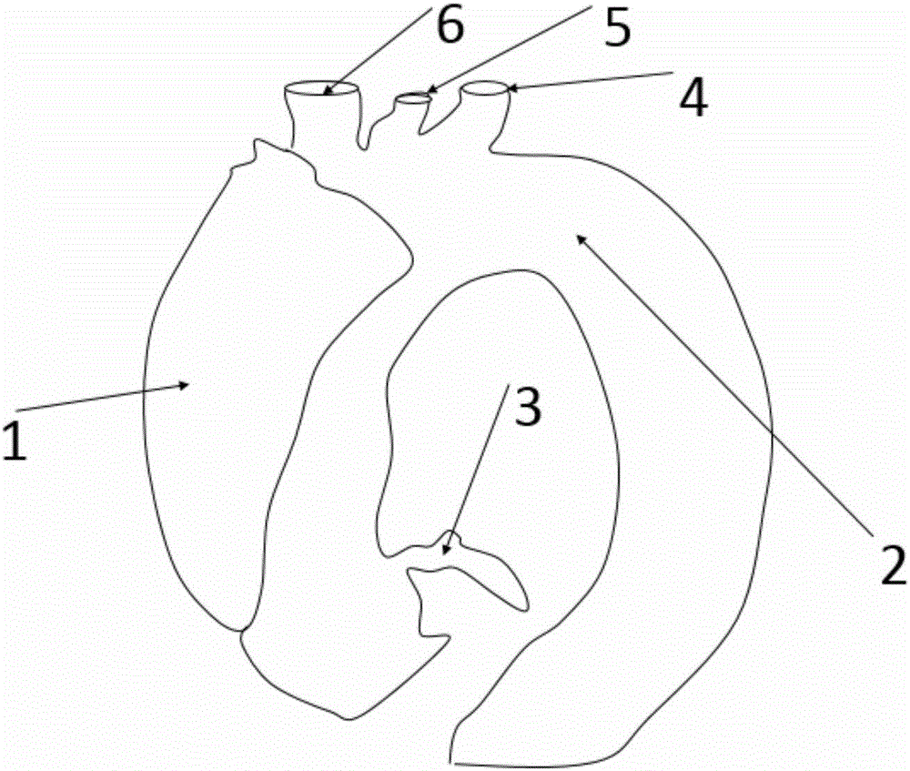
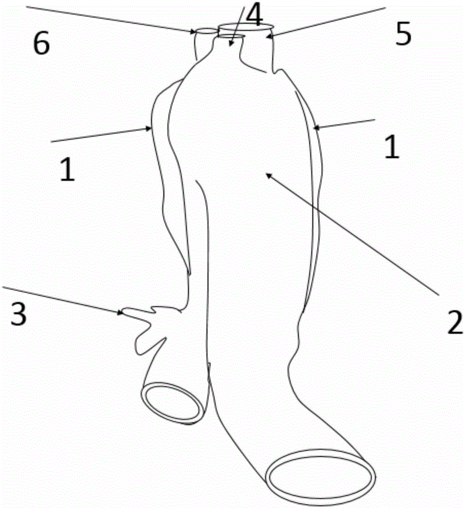
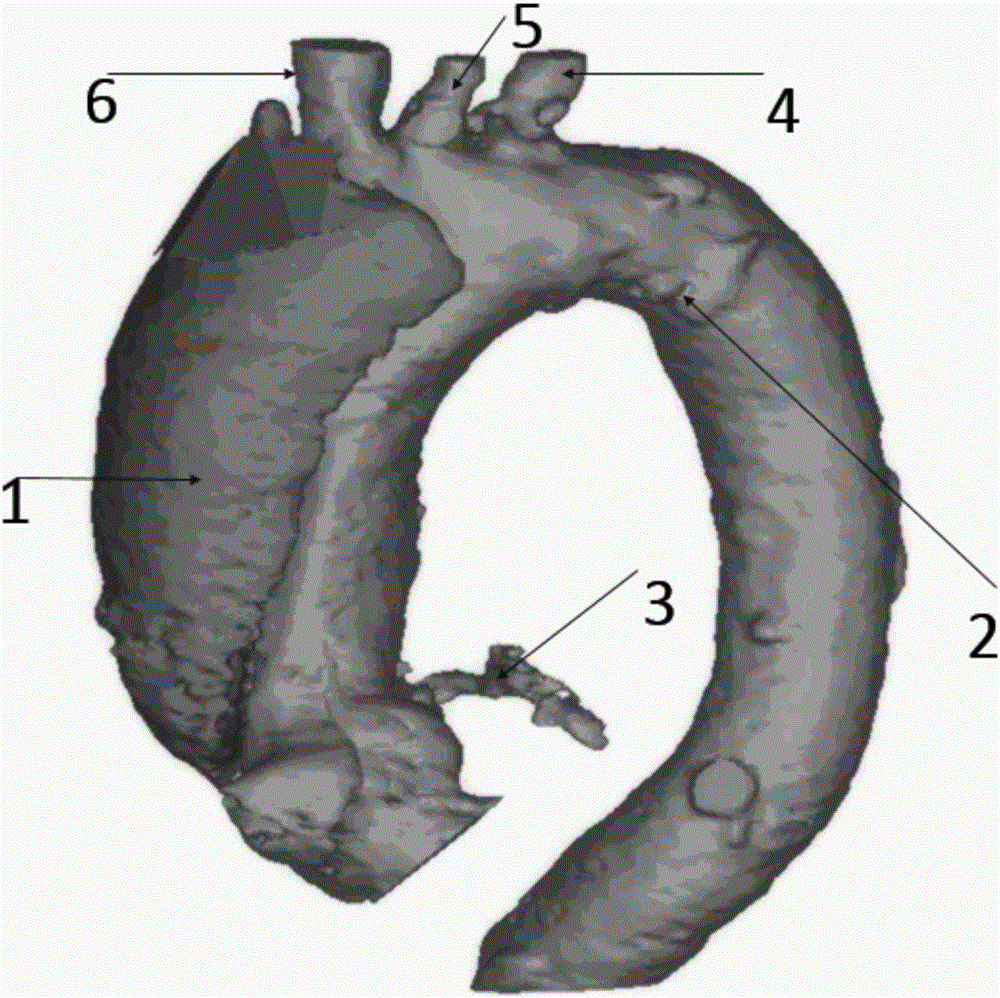
[0042] Example 1 as figure 1 , 2 , 3, an individualized aneurysm model and method thereof prepared using 3D printing technology, the specific method is as follows:

[0043] 1) Use CTA to perform thin-slice scanning of the chest vessels of patients with aortic arch aneurysms. The scanning range covers the patient's dissecting aneurysm 1, aortic arch 2, aortic arch 3 and its branches left subclavian artery 4, left common carotid artery 5, The brachiocephalic trunk 6 is used to obtain a DICOM format data file containing three-dimensional information data of the patient.

[0044] 2) Store the obtained image data in DICOM format on a CD or DVD disc, import it into Mimics 17.0 medical surgery design software (Materialise, Belgium), select the non-destructive format in the process of importing data, and perform three-dimensional images of regional blood vessels containing dissecting aneurysms modeling.

[0045] 3) Copy the created 3D image model to 3-Matic 9.0 (Materialise, Belgiu...

Embodiment 2

[0049] Example 2 as Figure 4 , 5 , 6, an individualized aneurysm model and method thereof prepared using 3D printing technology, the specific method is as follows:

[0050] 1) Using MRA to scan the cranial blood vessels of patients with intracranial aneurysm, the scanning range includes: internal carotid aneurysm 7 and internal carotid artery 8 . Obtain the DICOM format data file containing the three-dimensional information data of the patient.

[0051] 2) Store the obtained image data in DICOM format on a CD or DVD disc, import it into Mimics 17.0 medical surgery design software (Materialise, Belgium), select the non-destructive format in the process of importing data, and perform three-dimensional image construction on regional blood vessels containing aneurysms. model, including internal carotid aneurysm7 and internal carotid artery8.

[0052] 3) Copy the created 3D image model to 3-Matic 9.0 (Materialise, Belgium) software for outward expansion modeling with an expansion...


PUM

PropertyMeasurementUnit
Tube wall thicknessaaaaaaaaaa
Lengthaaaaaaaaaa

Abstract

The invention discloses an individual aneurysm model obtained through 3D printing technology and a manufacturing method thereof. The manufacturing method comprises the steps of firstly, performing high-precision layered scanning on an artery of an aneurysm patient by means of imaging equipment, thereby aneurysm image data; then performing sorting by means of inverse reconstruction software for obtaining the artery, and performing three-dimensional reconstruction; performing expansion amplification on the reconstructed artery by means of computer-aided 3D design software, then subtracting original blood vessels through Boolean operation, obtaining an empty aneurysm model and outputting a 3D printing identifiable file; and finally printing the aneurysm model by means of different materials through a 3D printer, wherein the size ratio between the aneurysm model and the aneurysm of the patient is 1:1. The aneurysm model obtained through the method can settle a defect of disease model shortage in China and can be applied in the fields of disease teaching, open heart surgery or interventional surgery training, surgery planning, etc. The aneurysm model and the manufacturing method have advantages of user-friendly customization, high accuracy, effective realizing of visuality and touchness, simple process, high forming speed and low cost.

Description

technical field [0001] The invention relates to the preparation of an aneurysm model for teaching and surgical training, in particular to an individualized aneurysm model based on 3D printing and a preparation method thereof. Background technique [0002] Aneurysm refers to local or diffuse abnormal expansion-like lesions of the arterial wall, often occurring in the ascending aorta, aortic arch, thoracic abdominal aorta, and intracranial arteries. Aneurysm rupture is one of the clinical critical illnesses. Aortic aneurysm rupture can cause shock in a short period of time, and the mortality rate is extremely high. The rupture of an intracranial aneurysm usually presents with severe headache as the first manifestation, and about 30% of patients may die outside the hospital due to secondary brain herniation within a short period of time. The diagnosis of aneurysm often requires the help of imaging examinations, common examination methods include digital subtraction angiography...

Claims


Application Information

Patent Timeline
IPC IPC(8): G09B23/28
CPCG09B23/28
Inventor 李翔宇
Owner 李翔宇
Learn More