
System and method for automatically detecting lesions in medical image through multi-model fusion

An automatic detection and medical image technology, which is applied in medical informatics, medical data mining, medical automatic diagnosis, etc., can solve problems such as lack, achieve the effect of improving accuracy, large theoretical value and economic benefits, and reducing false positives

Active Publication Date: 2017-05-17
周明
View PDF6 Cites 43 Cited by
  • Summary
  • Abstract
  • Description
  • Claims
  • Application Information

AI Technical Summary

Problems solved by technology

[0005] However, using deep learning technology to completely replace traditional computer-aided detection technology also has its shortcomings.
If a single deep learning strategy is used to realize computer-aided diagnosis, there will be a lack of comprehensive consideration of the combined use of various traditional computer-aided detection models and multiple detection models, and it may not be the optimal detection model solution.

Method used


Image

  • System and method for automatically detecting lesions in medical image through multi-model fusion
  • System and method for automatically detecting lesions in medical image through multi-model fusion
  • System and method for automatically detecting lesions in medical image through multi-model fusion

Examples


Embodiment Construction

[0045] The present invention will be described in further detail below in conjunction with the accompanying drawings and embodiments, and the embodiments are explanations of the present invention, rather than limitations.

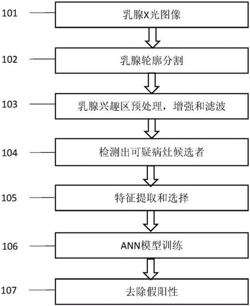
[0046] For the workflow of the existing breast CAD diagnosis system, please refer to figure 1 , each step listed in the figure is optimized separately in most cases, and each step passes the result as an input parameter to the subsequent step, with almost no feedback information. If an error occurred in the previous step, it will still be passed to the subsequent steps until the final result is obtained. Generally speaking, a mammogram 101 needs to go through breast contour segmentation 102, breast region of interest preprocessing 103, and detect suspicious lesions (lesions) candidates 104, after which processing, for example, feature extraction and selection 105 for the entire The performance of the system (sensitivity and specificity) plays the most import...


PUM


Abstract

The invention discloses a system and method for automatically detecting lesions in a medical image through multi-model fusion; the method comprises using single or fused detection models including deep learning technology to analyze and detect suspicious lesions in the medical image, such as a breast X-ray image. By using the system and method, lesion characteristics can be automatically extracted; the system and method are suitable for detecting or marking one or more types of lesions.

Description

technical field [0001] The present invention relates to a system and method for automatic detection of medical images combined with deep learning technology, in particular to using a single or fusion detection model including deep learning technology to detect suspicious lesions in medical images (such as mammograms) Systems and methods for detection and evaluation. Background technique [0002] Breast cancer is the most common cancer that threatens women's health. The key to preventing and treating breast cancer is early detection, early diagnosis and early treatment. Common modalities for breast health screening include X-rays, ultrasounds, and magnetic resonance imaging (MRI). Among them, mammography is considered the most accurate detection method, because it can detect various suspicious lesions (such as masses, microcalcifications, structural disorders, etc.) in the early stage. At present, mammogram diagnosis is mainly done by doctors visually, and the quality of d...

Claims


Application Information

Patent Timeline
Patent Type & Authority Applications(China)
IPC IPC(8): G06F19/00G06K9/32G06K9/46G06K9/62
CPCG16H50/20G16H50/70G06V10/25G06V10/44G06V2201/03G06F18/24
Inventor 劳志强张雪英周明
Owner 周明
Learn More