
Fusion protein for screening weak MdmX inhibitor or testing inhibition activity of weak MdmX inhibitor

A technology of fusion protein and inhibitory activity, which is applied in the field of biotechnology and can solve the problems of fluorescein losing its fluorescent function, reducing the accuracy of detection methods, and incompletely consistent molar ratios.

Active Publication Date: 2017-05-10
HUBEI UNIV OF TECH
View PDF2 Cites 2 Cited by
  • Summary
  • Abstract
  • Description
  • Claims
  • Application Information

AI Technical Summary

Problems solved by technology

[0010] 1. When the p53 fragment and the MdmX fragment are added to the test system separately, the molar ratio of the two is difficult to determine accurately, which will bring large systematic or random errors
[0011] 2. The protocol uses fluorescein to label p53 fragments, which will make the molar ratio of fluorescein molecules and p53 fragment molecules not completely consistent in each labeling operation, which will bring large random errors and make it more difficult to carry out accurate quantitative research
[0012] 3. In the protocol, fluorescein is used to label p53 fragments, and fluorescein is easy to decompose and denature under visible light and lose its fluorescence function, which requires corresponding operations to avoid light. Photolysis will reduce the accuracy of the detection method, and requiring light protection will increase the operation The difficulty and cost of experiments
[0013] 4. Due to the hydrophobicity of fluorescein, the accuracy of detection methods and data is reduced
[0014] 5. The p53 fragment and MdmX need to be prepared separately, which will increase the complexity of the operation
[0015] 6. Although MdmX and Mdm2 are homologous proteins and their three-dimensional structures are highly similar, the existing Mdm2 small molecule inhibitors have very weak inhibitory effect on MdmX
[0016] 7. The MdmX inhibitors with significantly different acting forces are all tested with the same model, and it is impossible to distinguish strong MdmX inhibitors from weak MdmX inhibitors. More importantly, weaker MdmX inhibitors may be due to ineffective Competitive binding to the p53 domain without detection

Method used


Image

  • Fusion protein for screening weak MdmX inhibitor or testing inhibition activity of weak MdmX inhibitor
  • Fusion protein for screening weak MdmX inhibitor or testing inhibition activity of weak MdmX inhibitor
  • Fusion protein for screening weak MdmX inhibitor or testing inhibition activity of weak MdmX inhibitor

Examples


Embodiment 1

[0071] Embodiment 1: p53p-XX-N-MdmX fusion protein expression plasmid construction

[0072] Based on the published crystal structure of the MdmX binding domain p53p of p53 and the p53 binding domain at the amino terminal of MdmX (PDB ID: 3DAB) (refer to Structure of the human Mdmx protein bound to the p53 tumor suppressor transactivation domain, Popowicz, G.M., Czarna , A., Holak, T.A. (2008) CellCycle 7:2441-2443), we established the following p53p-XX-N-MdmX fusion protein model.

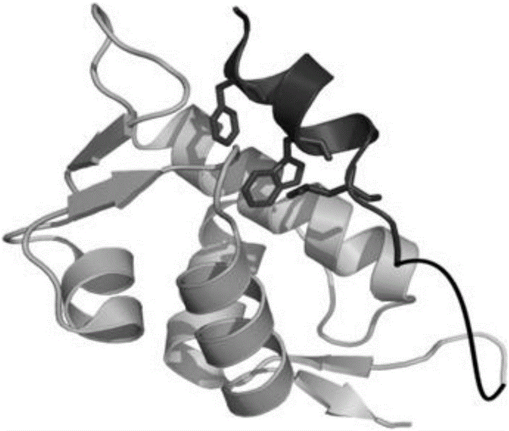
[0073] The amino acid sequence of the p53p-XX-N-MdmX fusion protein is shown below (the tertiary structure of the protein after removing the tag GSSHHHHHHGS is simulated by PymolWin as shown in figure 1 shown):

[0074] GSSHHHHHHGSSQETFSDLWKLLPENGSGSSENLYFQGSQINQVRPKLPLLKILHAAGAQGEMFTVKEVMHYLGQYIMVKQLYDQQEQHMVYCGGDLLGELLGRQSFSVKDPSPLYDMLRKNLVTLAT

[0075] Among them, SQETFSDLWKLLPEN is from the MdmX binding domain sequence of p53, GSQINQVRPKLPLLKILHAAGAQGEMFTVKEVMHYLGQYIMVKQLYDQQEQHMVYCGGDLLGELLG...

Embodiment 2

[0108] Example 2: Expression and purification of p53p-XX-N-MdmX fusion protein

[0109] Transform the pET28a-p53p-XX-N-MdmX plasmid into Escherichia coli BL21(DE3); pick a single colony into 2ml LB K + (Kanamycin) culture medium, 37°C, 200rpm for overnight culture; take 500μl bacterial solution and transfer it to 50ml LB K + Culture medium, 37°C, 200rpm for 3h; transfer 50ml of bacterial solution into 1L LB K + Culture medium, 37°C, 200rpm, until the cell concentration reaches OD 280nm =0.8 or so, add IPTG (final concentration 0.4mM) to induce, and collect the bacteria for about 20 hours (the centrifuge parameters are set to 3500rpm, 30min, 4°C when collecting the bacteria).

[0110] Ultrasonic cell disruptor disrupts cells. According to the ratio of cell volume:buffer volume=1:5, add bufferA buffer solution (50mM Na 2 HPO 4 , 200mM NaCl, 10mM imidazole, 1mM BME (β-mercaptoethanol), pH8.0), the parameters are set as: total time 2min; ultrasonic time 2s; interval time 4s; ...

Embodiment 3

[0112] Embodiment 3: Fluorescence spectrum analysis of p53p-XX-N-MdmX fusion protein model

[0113] Take a frozen 200μl p53p-XX-N-MdmX protein, thaw it quickly, and dilute the protein concentration to OD with phosphate buffer 280nm = 0.1. Take 400 μl to a quartz cuvette, and perform fluorescence scanning on a F-7000 fluorescence spectrophotometer. According to the endogenous fluorescence characteristics of tryptophan, take λ EX 278nm, scan λ EM In the fluorescence intensity in the range of 290nm~500nm, it was found that the maximum fluorescence intensity was at 321nm ( Figure 4 a). Therefore, λ EM 321nm, scan λ EX It is the fluorescence excitation spectrum curve of 245nm~300nm, the result is as follows Figure 4 As shown in b, it can also be known that at λ EX At 278nm, the fluorescence intensity is the largest, so the wavelength of excitation light is set at 278nm.


PUM


Abstract

The invention discloses a fusion protein for screening a weak MdmX inhibitor or testing the inhibition activity of a weak MdmX inhibitor. The testing sequence is an MdmX sequence or a homologous sequence thereof. The combination sequence is the mutant of p53 segment of the MdmX binding domain of p53 sequence. The linker arm sequence is a peptide segment, and the upstream and lower stream of the linker arm sequence are connected to one of the testing sequence and the combination sequence. The emission spectrums of tryptophan in the fusion protein are different when the combination structural domain and the testing structural domain are connected / disconnected; and for the mutant of p53 segment, compared with a wild p53 segment, the interaction between the p53 combination structural domain and the MdmX combination structural domain is weakened, so that the MdmX inhibitor with a weak binding force can compete with the combination sequence to combine the testing sequence, and thus the inhibition degree of the weak MdmX inhibitor on MdmX can be effectively measured.

Description

technical field [0001] The invention belongs to the field of biotechnology, and relates to the preparation and application of a recombinant protein for screening MdmX inhibitors or testing the inhibitory activity of Mdmx inhibitors, in particular to screening for weak MdmX inhibitors or testing the inhibition of weak Mdmx inhibitors The preparation and application of an active recombinant protein, more specifically relates to the preparation of a p53 polypeptide and MdmX amino-terminal fusion protein and its analogue and its application in screening MdmX inhibitors or testing the inhibitory activity of MdmX inhibitors. Background technique [0002] p53 is a tumor suppressor protein that plays a role in biological processes such as cell cycle arrest, DNA damage repair and apoptosis, and has become an important target for tumor therapy. The anti-oncogenic activity of p53 is suppressed by intracellular overexpression of MdmX and Mdm2, which is associated with more than half of ...

Claims


Application Information

Patent Timeline
IPC IPC(8): C07K19/00C12N15/62C12N15/70
CPCC12N15/70C07K14/4702C07K14/4746C07K2319/00C12N2800/101Y02A50/30
Inventor 苏正定阳飞张华山成细瑶
Owner HUBEI UNIV OF TECH
Learn More