
Two-pass region growing and morphological reconstruction combination-based lung airway tree segmentation method

A region growing and morphological technology, applied in the field of medical image processing, can solve the problems of less advanced peripheral trachea, high complexity, and too long time, and achieve the effect of increasing the number of branches of the trachea tree and being simple and easy to implement

Active Publication Date: 2016-11-09
UNIV OF SHANGHAI FOR SCI & TECH +1
View PDF5 Cites 21 Cited by
  • Summary
  • Abstract
  • Description
  • Claims
  • Application Information

AI Technical Summary

Problems solved by technology

[0003] The paper "Vessel-guided airway tree segmentation: A voxel classification approach" published by Lo et al. in the Journal of Medical Image Analysis in 2010 proposed a tracheal segmentation method based on pulmonary vascular guidance. This method uses the parallel anatomical structure of blood vessels and trachea Features, using blood vessels as a segmentation guide, can more accurately segment the pulmonary trachea tree, but the algorithm needs to determine the direction of blood vessels, which is complex and takes too long; KaiLAI et al. published a paper at the International Conference on Bioinformatics and Biomedical Engineering in 2009 "Automatic 3D Segmentation of Lung Airway Tree: A NovelAdaptive Region Growing Approach" proposes a segmentation method based on the division of the region of interest. Although the method of first dividing the region of interest and then segmenting it can effectively prevent the segmentation from leaking to the lung parenchyma, and at the same time Improve the efficiency of the algorithm, but the experimental results show that the detected high-level peripheral trachea is less, and the segmentation effect is not good; Michael W et al published the paper "Robust 3-D airway treesegmentation for image-guided" in the journal IEEE Transactions on Medical Imaging in 2010 "peripheral bronchoscopy" combines region growth with tracheal geometric structure analysis, and uses local image features as a guide to segment the pulmonary trachea tree. Although this method can detect more advanced peripheral trachea and has better accuracy, it is vulnerable to Influenced by factors such as noise and artifacts in low-dose CT images, and there are too many parameters that need to be manually set; Anna et al. published a paper "Two-pass region growing algorithm for segmenting airway from MDCT chest scans" uses the double-pass region growing method to detect small trachea through morphological erosion and expansion operations, but the detection effect on advanced peripheral trachea is not good, and the performance is poor on CT images with low dose and thick slice thickness. The branches of the trachea at all levels are quantitative indicators, and the tenth-level trachea can be segmented at most. The total number of trachea branches is at most 145, and the segmentation effect needs to be improved

Method used


Image

  • Two-pass region growing and morphological reconstruction combination-based lung airway tree segmentation method
  • Two-pass region growing and morphological reconstruction combination-based lung airway tree segmentation method
  • Two-pass region growing and morphological reconstruction combination-based lung airway tree segmentation method

Examples


Embodiment

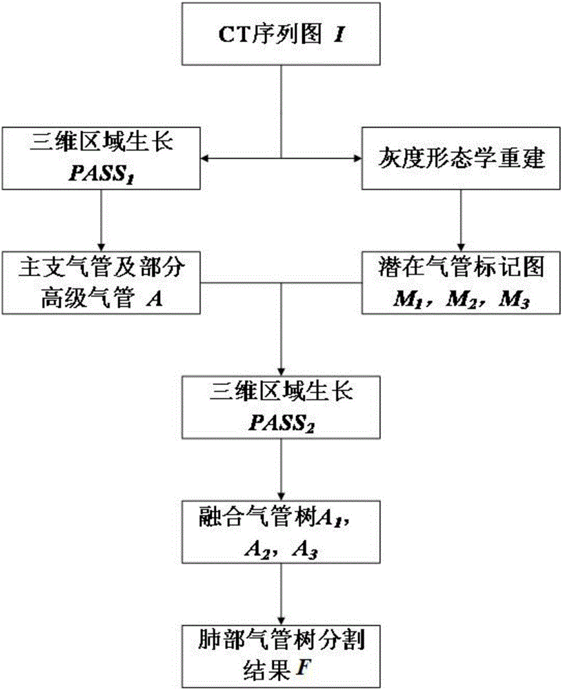
[0032] figure 1 It is a method flow chart of the pulmonary tracheal tree segmentation method of double-stroke region growing combined with morphological reconstruction in the embodiment of the present invention; figure 2 It is a segmentation path diagram of the pulmonary tracheal tree segmentation method based on double-pass region growing combined with morphological reconstruction in the embodiment of the present invention.

[0033] like figure 1 As shown, in the embodiment of the present invention, the pulmonary tracheal tree segmentation method of double-stroke region growing combined with morphological reconstruction includes the following steps:

[0034] Step 1, input the sequential tomographic images of chest CT in DICOM format to be segmented.

[0035] Step 2, through automatic selection, the three-dimensional region growth seed point P1 is obtained in the sequential tomographic image. The CT value of the inside of the pulmonary trachea ranges from -1024HU to -800HU...


PUM


Abstract

The invention aims to provide a two-pass three-dimensional region growing and morphology combination-based lung airway tree segmentation algorithm for solving the problem to realize effective segmentation of a lung airway tree. The two-pass region growing and morphological reconstruction combination-based lung airway tree segmentation method provided by the invention comprises the following steps of automatically obtaining a seed point P1 of three-dimensional region growing from a serial section image; performing first-pass region growing by taking P1 as the seed point to obtain a lung main airway tree; setting a multi-scale morphological operator, and obtaining corresponding airway marking graphs in three anatomic positions of a cross section, a coronal plane and a sagittal plane by utilizing gray-scale morphological reconstruction operation; performing second-pass region growing by taking the lung main airway tree as a basis and the airway marking graphs in the three anatomic positions as limitation conditions to obtain corresponding segmented airway trees; and fusing the segmented airway trees to obtain a final lung airway tree segmentation result F.

Description

technical field [0001] The invention relates to the field of medical image processing, in particular to a pulmonary tracheal tree segmentation method based on double-stroke region growing combined with morphological reconstruction. Background technique [0002] The anatomical structure of the pulmonary tracheal tree is the pathological criterion for diagnosing various lung diseases such as bronchial stenosis, emphysema, and COPD. Therefore, whether the pulmonary tracheal tree can be accurately segmented from CT images will ultimately affect to the accuracy of clinical diagnosis. Pulmonary trachea is an important tissue for gas exchange between the human body and the outside world. It extends from the throat to the main bronchus in sequence, forming a tree-like structure with complex shapes and variable directions. Due to the partial volume effect in the CT image, the motion artifact caused by the movement of the patient during the shooting of the CT image, and the limitatio...

Claims


Application Information

Patent Timeline
IPC IPC(8): G06T7/00
CPCG06T7/0012G06T2207/10081G06T2207/30061
Inventor 段辉宏陈轶群聂生东龚敬刘霁雨贺兴怡代晓婷
Owner UNIV OF SHANGHAI FOR SCI & TECH
Learn More