
Method for non-invasive measurement of right ventricular pressure and pulmonary artery pressure method

A technology for pulmonary artery pressure and ventricular pressure, which is applied in the field of medical imaging examination to achieve low examination costs and real-time monitoring

Inactive Publication Date: 2016-09-21
SHANGHAI FIRST PEOPLES HOSPITAL
View PDF0 Cites 2 Cited by
  • Summary
  • Abstract
  • Description
  • Claims
  • Application Information

AI Technical Summary

Problems solved by technology

Currently, noninvasive methods for right ventricular and pulmonary artery pressures have not been reported

Method used


Image

  • Method for non-invasive measurement of right ventricular pressure and pulmonary artery pressure method
  • Method for non-invasive measurement of right ventricular pressure and pulmonary artery pressure method
  • Method for non-invasive measurement of right ventricular pressure and pulmonary artery pressure method

Examples


Embodiment Construction

[0025] The specific embodiments provided by the present invention will be described in detail below in conjunction with the accompanying drawings.

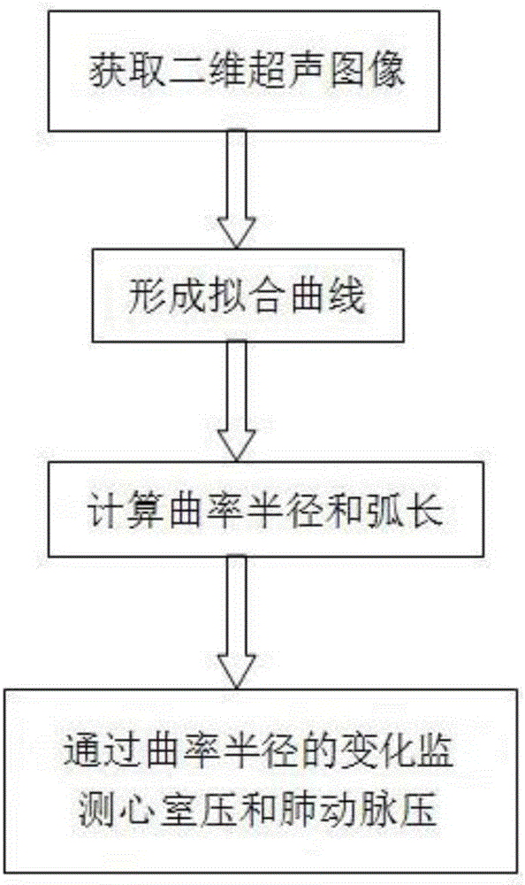
[0026] as attached figure 1 As shown, a method for non-invasive detection of right ventricular pressure and pulmonary artery pressure, comprising the following steps:

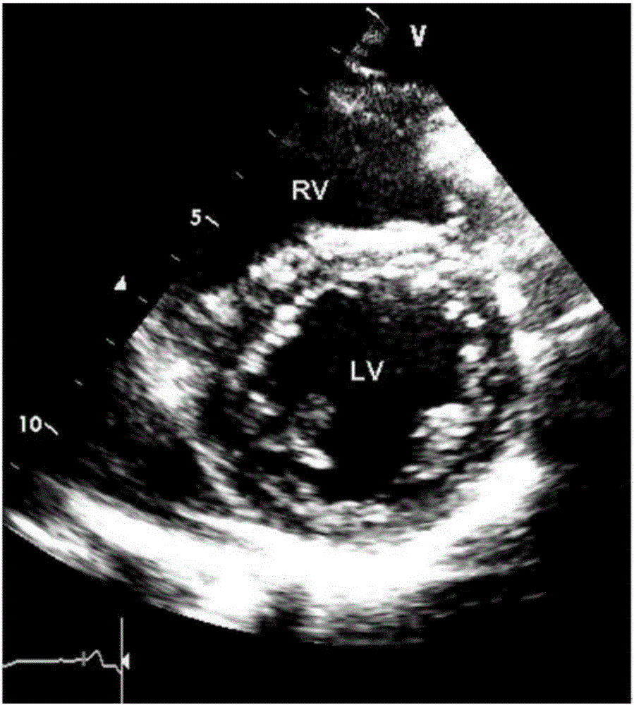
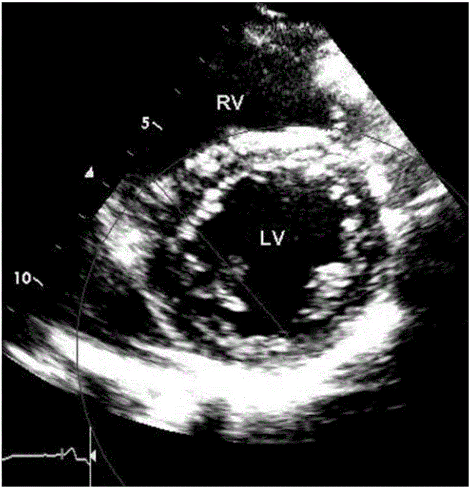
[0027] a. In left ventricular short-axis view, store the end-systolic and end-diastolic short-axis two-dimensional ultrasound images of the ventricle at the level of the mitral valve, and the two-dimensional ultrasound images of the end-diastole at the level of the mitral valve chordae during inspiratory phase and expiratory phase (if attached figure 2 shown);

[0028] b. Draw points on the interventricular septum or wall of the above image to form a fitting curve of an arc-shaped interventricular septum or wall, and calculate the radius of curvature and arc length of the fitting curve;

[0029] c. The tubular hypothetical formula combined with Laplace’s theore...


PUM


Abstract

The invention relates to a method for non-invasive measurement of right ventricular pressure and pulmonary artery pressure method. The method comprises the following steps of obtaining a bicuspid valve horizontal left ventricle short-axis two-dimensional ultrasonic image; drawing points and interventricular septum, a fitting curve of an arc-shaped interventricular septum is formed, the curvature radius and an arc length of the fitting curve are worked out; on the basis of the tubular hypothesis formula of the laplace's theorem, and the left and right ventricle wall pressure is obtained through calculation. Accordingly, non-invasive measurement, ventricular pressure monitoring and pulmonary artery pressure monitoring can be achieved by researching the ventricular wall curvature changes of left and right ventricles.

Description

【Technical field】 [0001] The invention relates to the technical field of medical image inspection applications, in particular to a method for non-invasive detection of right ventricular pressure and pulmonary artery pressure. 【Background technique】 [0002] The onset of right heart failure and acute and chronic pulmonary hypertension is hidden and difficult to detect. Once obvious clinical symptoms appear, the disease progresses rapidly, the prognosis is poor, and the mortality rate is high. In particular, patients with acute pulmonary embolism have a higher mortality rate. Accurate assessment of pulmonary artery pressure and right ventricular pressure in such patients is a race against time, and clinical intervention needs to be done as soon as possible. Currently, the gold standard for measuring right ventricular pressure and pulmonary artery pressure is cardiac catheterization and pulmonary artery catheterization. This method is invasive, expensive, and cannot be reuse...

Claims


Application Information

Patent Timeline
IPC IPC(8): A61B8/00A61B8/04
CPCA61B8/04A61B8/52A61B8/5223
Inventor 罗向红李朝军
Owner SHANGHAI FIRST PEOPLES HOSPITAL
Learn More