Method for displaying human arginase1 on surfaces of escherichia coli

An arginase and Escherichia coli technology, applied in the surface field of human arginase-1, can solve the problems of carrying various viruses, long application time, complicated preparation process, etc. The effect of reducing synthesis cost and simplifying purification steps

Inactive Publication Date: 2016-08-24
HUBEI UNIV
View PDF2 Cites 8 Cited by
  • Summary
  • Abstract
  • Description
  • Claims
  • Application Information

AI Technical Summary

Problems solved by technology

[0003] Arginase is difficult to achieve high-efficiency soluble expression in Escherichia coli, and it is isolated and extracted from the liver, which is the main source of arginase, but there is a risk of carrying various viruses
Human arginase-1 has been secreted and expressed in Pichia pastoris, and the purified human arginase-1 can effectively transform L-Arg into L-Orn after chitin entrapment However, this method has the disadvantages of long time use, complicated preparation process, and easy loss of enzyme activity during operation.

Method used


Image

  • Method for displaying human arginase1 on surfaces of escherichia coli
  • Method for displaying human arginase1 on surfaces of escherichia coli
  • Method for displaying human arginase1 on surfaces of escherichia coli

Examples


Embodiment 1

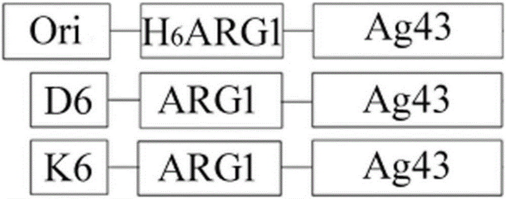
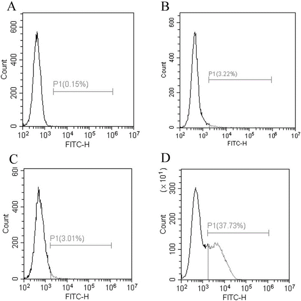
[0028] The method of the present invention is used to display human arginase-1 in Escherichia coli. First, the constructed recombinant plasmid pET23a / H 6ARG1-Ag43 transformed Escherichia coli competent cell Rosetta Blue strain, and cultured overnight at 37°C. Then, pick a single colony and inoculate it in 100mL LB medium. The final concentration of antibiotic ampicillin is 100μg / mL. Shake culture at 37°C. The OD value is between 0.5 and 0.6. Incubate for 8 hours. Then, the cells were collected by centrifugation at 12000 RPM at 4°C, washed with pre-cooled PBS for 3 times, and finally resuspended in 10 mL of pre-cooled PBS buffer for later use. Then it was measured by Chinard reaction and converted to the corresponding human arginase-1 enzyme activity. ( picture 3C , picture 4 )

Embodiment 2

[0030] The method of the present invention is used to display human arginase-1 in Escherichia coli. First, the constructed recombinant plasmid pET23a / H 6 D. 6 - ARG1-Ag43 was transformed into Escherichia coli competent cell Rosetta Blue strain, and cultured overnight at 37°C. Then, pick a single colony and inoculate it in 100mL LB medium, the final concentration of antibiotic ampicillin is 100μg / mL, shake culture at 37°C, the OD value is between 0.5 and 0.6, add IPTG, the final concentration is 1mM, 37°C Induction culture was carried out for 8 hours. Then, the cells were collected by centrifugation at 12000 RPM at 4°C, washed with pre-cooled PBS for 3 times, and finally resuspended in 10 mL of pre-cooled PBS buffer for later use. Then it was measured by Chinard reaction and converted to the corresponding human arginase-1 enzyme activity. ( picture 3B , picture 4 )

Embodiment 3

[0032] The method of the present invention is used to display human arginase-1 in Escherichia coli. First, the constructed recombinant plasmid pET23a / H 6 K 6 - ARG1-Ag43 was transformed into Escherichia coli competent cell Rosetta Blue strain, and cultured overnight at 37°C. Then, pick a single colony and inoculate it in 100mL LB medium. The final concentration of antibiotic ampicillin is 100μg / mL. Shake culture at 37°C. The OD value is between 0.5 and 0.6. Incubate for 8 hours. Then, the cells were collected by centrifugation at 12000 RPM at 4°C, washed with pre-cooled PBS for 3 times, and finally resuspended in 10 mL of pre-cooled PBS buffer for later use. Then it was measured by Chinard reaction and converted to the corresponding human arginase-1 enzyme activity. ( picture 3D , picture 4 )

[0033] Various example result analysis

[0034] After flow cytometry analysis, the charged polypeptide 6×Glu and 6×Lys can improve the display efficiency of Antigen 43 protein...


PUM


Abstract

The invention provides a method for displaying human arginase1 on surfaces of escherichia coli. The method comprises steps as follows: 1) recombinant plasmids for surface display of human arginase1 are constructed; 2) the recombinant plasmids are converted into escherichia coli competent cells, and genetic engineering strains are obtained; 3) the recombinant strains are subjected to shake-flask culture, and the display efficiency and enzyme activity of human arginase1 are detected; 4) L-Arginine (L-arginine, L-Arg) is efficiently converted by means of the recombinant strains for synthesis of L-Ornithine (L-ornithine, L-Orn). Human arginase1 is effectively displayed on surfaces of escherichia coli cells through improved Type V self-transport protein (Antigen 43), and industrial application of human arginase1 is realized more rapidly. By comparison with an original Type V self-transport protein (Antigen 43) display system, the display efficiency and the enzyme activity of human arginase1 are remarkably improved; by comparison with an existing chitin immobilization method, the synthesis cost is reduced, the technological process is shortened, and the purification procedures are simplified.

Description

technical field [0001] The present invention relates to the surface technology of human arginase-1 (Human Arginase 1), especially through the optimized autotransporter Antigen 43 surface display method, which is a new idea of ​​human arginase I display, New ways, new methods. Background technique [0002] Human arginase-1 exists in human liver cells, participates in the urea metabolic cycle, and catalyzes the hydrolysis of L-Arg to generate L-Orn and urea. Arginase I deficiency can cause a series of related diseases, such as developmental delay, mental retardation, epilepsy and movement disorders. Human arginase-1 is also a potential clinical drug, which can be used to treat cancer. [0003] Arginase is difficult to achieve high-efficiency soluble expression in Escherichia coli, and it is isolated and extracted from the liver, which is the main source of arginase, but there is a risk of carrying various viruses. Human arginase-1 has been secreted and expressed in Pichia p...

Claims


Application Information

Patent Timeline
IPC IPC(8): C12N9/78C12N15/70C12N1/21C12R1/19
CPCC12N9/78C12Y305/03001
Inventor 张贞马立新卞璐唐荣兴
Owner HUBEI UNIV
Why Eureka
Try Eureka