
Detection and analysis method for thermodynamic parameters of medulloblastoma cells and purpose thereof

A technology of medulloblastoma and analysis method, applied in the direction of material thermal development, etc., can solve the problem of not being able to find the M1 state of medulloblastoma, and achieve optimized purification procedures, improved sensitivity and specificity, and low interference factors Effect

Active Publication Date: 2016-08-03
AFFILIATED HUSN HOSPITAL OF FUDAN UNIV
View PDF1 Cites 2 Cited by
  • Summary
  • Abstract
  • Description
  • Claims
  • Application Information

AI Technical Summary

Problems solved by technology

[0003] Usually, in clinical practice, the presence of tumor cells is mainly identified through imaging and tumor exfoliation cytology. Among them, imaging diagnosis is still the main standard, especially when detecting the presence of medulloblastoma cells, it can be more objectively displayed. The presence of medulloblastoma cells, that is, imaging methods can provide a reference for clinical detection of MB intracranial or extracranial metastatic lesions, but it still requires experienced neurosurgeons or imaging doctors to determine Comprehensive judgment, but it can not find the M1 status of medulloblastoma, and the M1 status is also an important indicator affecting the survival time of patients, and currently the M1 status can only be checked by exfoliative cytology, but unfortunately, exfoliative cytology does not matter There are deficiencies in both specificity and sensitivity. For example, in terms of specificity, exfoliated cytology is strictly limited by the conditions and time of lumbar puncture examination. The exfoliated tumor cells found in the early postoperative period do not necessarily indicate the specificity of tumor cells. In terms of sensitivity, the sensitivity of exfoliative cytology is only about 35% (TerterovS, JNeurosurgPediatr, 2010)

Method used


Image

  • Detection and analysis method for thermodynamic parameters of medulloblastoma cells and purpose thereof
  • Detection and analysis method for thermodynamic parameters of medulloblastoma cells and purpose thereof
  • Detection and analysis method for thermodynamic parameters of medulloblastoma cells and purpose thereof

Examples


Embodiment 1

[0039] Example 1 Detection and analysis experiment of medulloblastoma cell thermodynamic parameters

[0040] 1. Sample processing of the cerebrospinal fluid (CSF) of the isolated patient:

[0041] 1) Take CSF samples stored at -80°C and thaw naturally at room temperature;

[0042] 2) Load the sample into an AmiconUltra-0.5ml ultracentrifuge tube, cap each sample tube and mark it according to the sample number;

[0043] 3) Ultrafiltration and centrifugation at 14000×g at 4°C for 30 minutes;

[0044] 4) Pour off the liquid in the bottom collection tube, add 450ul 4°C pre-cooled PBS (PH=7.4) to the filter element, and centrifuge at 14000×g for 30 minutes;

[0045] 5) Pour off the liquid in the bottom collection tube, add 450ul 4°C pre-cooled PBS (PH=7.4) to the filter element, and centrifuge at 14000×g for 30 minutes;

[0046] 6) After the last round of centrifugation, take out the filter element, place it upside down on another clean collection tube, and centrifuge at 1000×g ...


PUM

PropertyMeasurementUnit
Sensitivityaaaaaaaaaa

Abstract

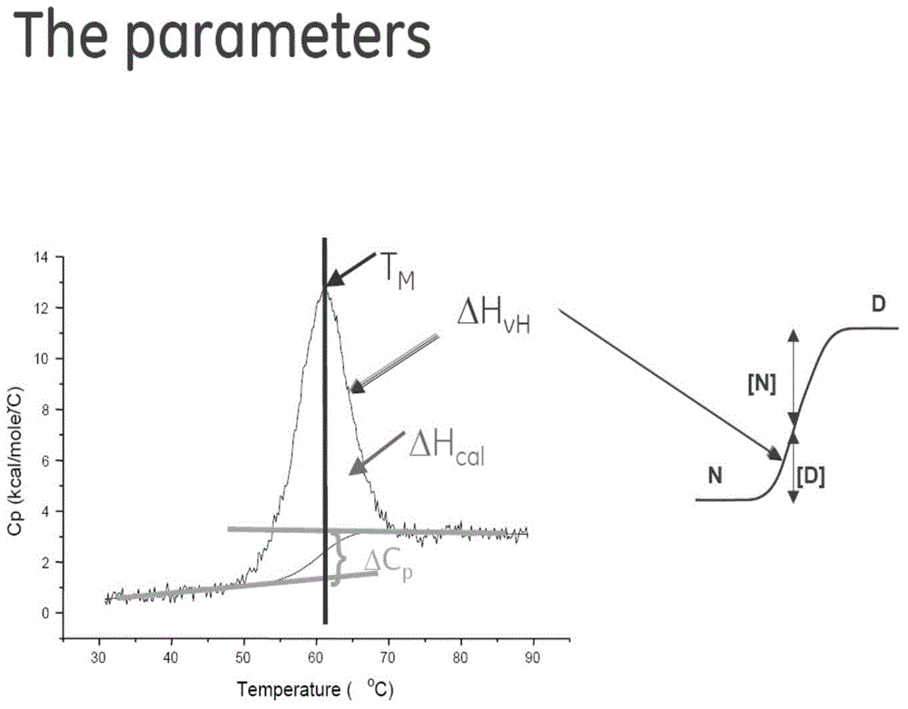
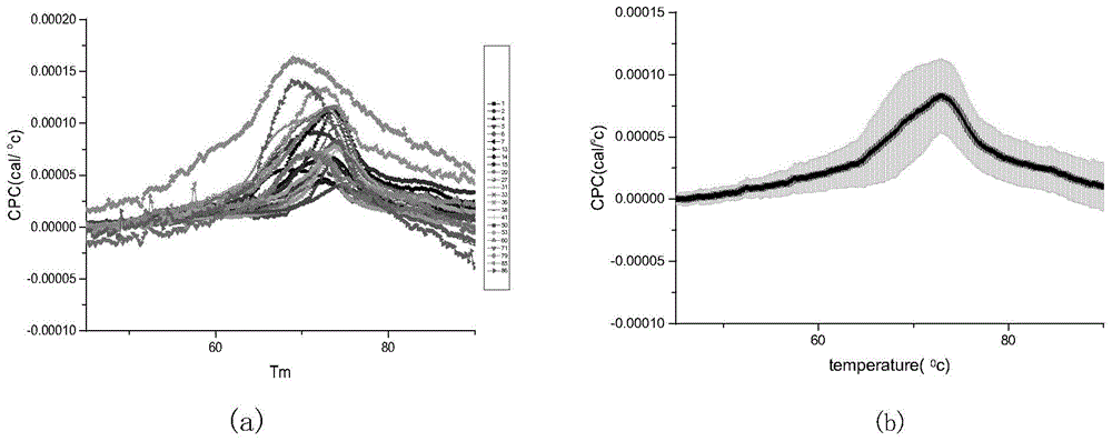
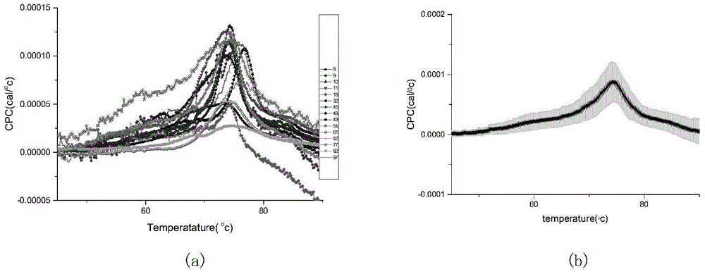
The invention belongs to the technical field of clinical examination, and relates to a detection and analysis method for thermodynamic parameters of medulloblastoma cells and purpose thereof. According to the method, through a differential scanning calorimetry (DSC), an in-vitro patient cerebrospinal fluid (CSF) specimen is heated, thermodynamic parameter variation generated during a heating process is analyzed, and then a parameter critical value is determined. The method optimizes a purification treatment process of cerebrospinal fluid protein, experiment operation is simplified, the CSF specimen Tm can be directly and rapidly detected, when Tm is 73.76 DEG C, the specificity is 69.6%, sensitivity is 86.7%, and the value is the detection threshold value which is an auxiliary determination for dissemination and transfer of the medulloblastoma cells. No expensive experiment equipment is required in the method, and the method has the advantages of simple and easy operation, and provides favorable tools for researches of dissemination and transfer behaviors of the medulloblastoma cells.

Description

technical field [0001] The invention belongs to the technical field of clinical testing and relates to the detection of medulloblastoma cells, in particular to a method for detecting and analyzing thermodynamic parameters of medulloblastoma cells and its application. In the method of the present invention, differential scanning calorimetry (differential scanning calorimetry, DSC) is used to heat the obtained isolated patient's cerebrospinal fluid (Cerebrospinal fluid, CSF) specimen, and the thermodynamic parameter changes generated during the heating process are analyzed to determine the critical value of the parameter, The method of the invention and the obtained parameters are helpful for judging the dissemination and metastasis of medulloblastoma. Background technique [0002] It has been reported in the literature that medulloblastoma (M) occurs frequently in children. This tumor originates from embryonic residual cells and has a short survival time. It is one of the mos...

Claims


Application Information

Patent Timeline
Patent Type & Authority Applications(China)
IPC IPC(8): G01N25/20
Inventor 姚瑜唐超周良辅张振宇沈方钟平
Owner AFFILIATED HUSN HOSPITAL OF FUDAN UNIV
Learn More