
Partition method for kidney artery CT contrastographic picture vessels based on three-dimensional Zernike matrix

A technology of contrast images and blood vessels, which is applied in the field of blood vessel segmentation and image processing of renal arterial CT contrast images, can solve the problems of over-segmentation and under-segmentation of blood vessels, and achieve the effect of solving under-segmentation and over-segmentation and ensuring segmentation accuracy

Inactive Publication Date: 2016-07-20
SOUTHEAST UNIV
View PDF3 Cites 5 Cited by
  • Summary
  • Abstract
  • Description
  • Claims
  • Application Information

AI Technical Summary

Problems solved by technology

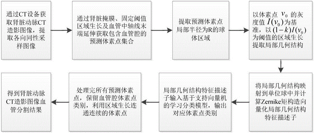
[0021] Purpose of the invention: In order to effectively solve the problem of over-segmentation and under-segmentation of blood vessels encountered by the threshold region growing method in the blood vessel segmentation of CT angiography images of renal arteries, the present invention provides a blood vessel segmentation method for CT angiography images of renal arteries based on three-dimensional Zernike moments , this method is based on the difference in the local geometric structure of the vascular lumen and other tissue voxel points in the arterial CT angiography image, by calculating the three-dimensional Zernike moment to construct the local geometric structure feature descriptor vectorization to characterize these geometric structure features, and through the support vector machine The learning classification model is used to classify, and then obtain the voxel points of the vascular cavity

Method used


Image

  • Partition method for kidney artery CT contrastographic picture vessels based on three-dimensional Zernike matrix
  • Partition method for kidney artery CT contrastographic picture vessels based on three-dimensional Zernike matrix
  • Partition method for kidney artery CT contrastographic picture vessels based on three-dimensional Zernike matrix

Examples


Embodiment Construction

[0048] The present invention will be further described below in conjunction with the accompanying drawings.

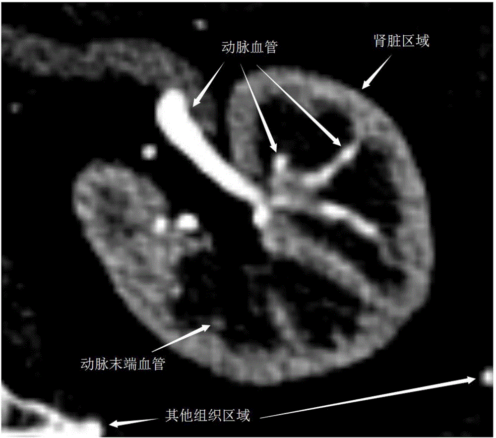
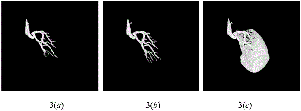
[0049] Affected by the contrast agent, the blood vessels in the renal arterial CT angiography image will show a higher CT value (such as figure 1 shown). Due to differences in image acquisition machines, measurement of contrast injections, and human body structure, the CT value of terminal arteries will be relatively low, and it will be relatively close to the CT value of surrounding tissues. On the other hand, kidney stones will also exist near blood vessels. The method of obtaining arterial vessels caused by threshold area growth is troublesome, that is, the problem of over-segmentation and under-segmentation of blood vessels (such as image 3 shown). The threshold is not fixed and needs to be fine-tuned, which greatly reduces the doctor's work efficiency. At the same time, the segmentation of the end of the blood vessel obtained by region growing is not accurate. ...


PUM


Abstract

The invention discloses a partition method for kidney artery CT contrastographic picture vessels based on a three-dimensional Zernike matrix. The partition method comprises the following steps: firstly, acquiring a forecast voxel point set and extracting a local geometric structure of the voxel point; then, constructing a descriptor with space rotation invariance by mapping the local geometric structure into a unit ball and solving the three-dimensional Zernike matrix; using the local geometric structure characteristic descriptors composed of different order and repeatability descriptors for expressing the characteristics of the local geometric structure in a quantizing form; and finally, adopting a study classifying model based on a support vector machine for classifying the acquired local geometric structure characteristic descriptors, thereby confirming the voxel points in the vessel lumen and acquiring a final vessel partition result through region growth. According to the method provided by the invention, the semiautomatic and accurate partition for kidney artery CT contrastographic picture vessels is realized, and the working efficiency of doctors and the accuracy of clinic auxiliary diagnosis are promoted.

Description

technical field [0001] The invention relates to a three-dimensional Zernike moment-based blood vessel segmentation method for renal arterial CT angiography images, which belongs to the image processing technology. Background technique [0002] Vascular disease has become one of the most important public health problems. Faced with specific size and complex angiographic images, segmenting blood vessels from them is a key step towards accurate visualization, diagnosis and quantitative analysis of vascular lesions. Among them, renal artery-related diseases such as arterial stenosis and renal tumors have become the main reasons for the need for renal artery segmentation. Renal arterial stenosis can cause renovascular hypertension and renal dysfunction, and renal tumor resection requires ligation of the branches of blood vessels in the artery, all of which require precise segmentation of renal arteries. [0003] CT angiography (Computed Tomographic Angiography, CTA) is a non-inv...

Claims


Application Information

Patent Timeline
IPC IPC(8): G06T7/00
CPCG06T2207/10081G06T2207/20081G06T2207/30084
Inventor 杨冠羽丁皓顾金金舒华忠
Owner SOUTHEAST UNIV
Learn More