
In-vitro culture method of vaginal epithelial cells of mouse

A technology for vaginal epithelial cells and in vitro culture, applied in cell dissociation methods, reproductive tract cells, artificial cell constructs, etc., can solve the problems of long passage cycle, obtaining a large number of high-purity seed cells, and obtaining a small number of cells, etc. To achieve the effect of convenient operation

Inactive Publication Date: 2016-03-30
祁文瑾
View PDF1 Cites 2 Cited by
  • Summary
  • Abstract
  • Description
  • Claims
  • Application Information

AI Technical Summary

Problems solved by technology

[0002] For the in vitro culture of mouse vaginal epithelial cells, domestic and foreign studies have used the tissue block method to culture vaginal epithelial cells. Although the operation is simple, it takes a long time and the passage cycle is long
[0003] In recent years, the mouse vaginal epithelium was isolated by enzymatic digestion, and the epithelial layer was digested into a single cell suspension by enzymatic digestion. After successful culture, it was used for tissue engineering vaginal research, but due to the However, due to the small number of cells obtained, the inability to adhere to the wall, and the poor ability of passage, there has not been a large number of high-purity seed cells obtained in a relatively short period of time, which can be used to observe the physiological and pathological changes of the vaginal epithelium, the process of carcinogenesis, and organize Construction of the project

Method used


Image

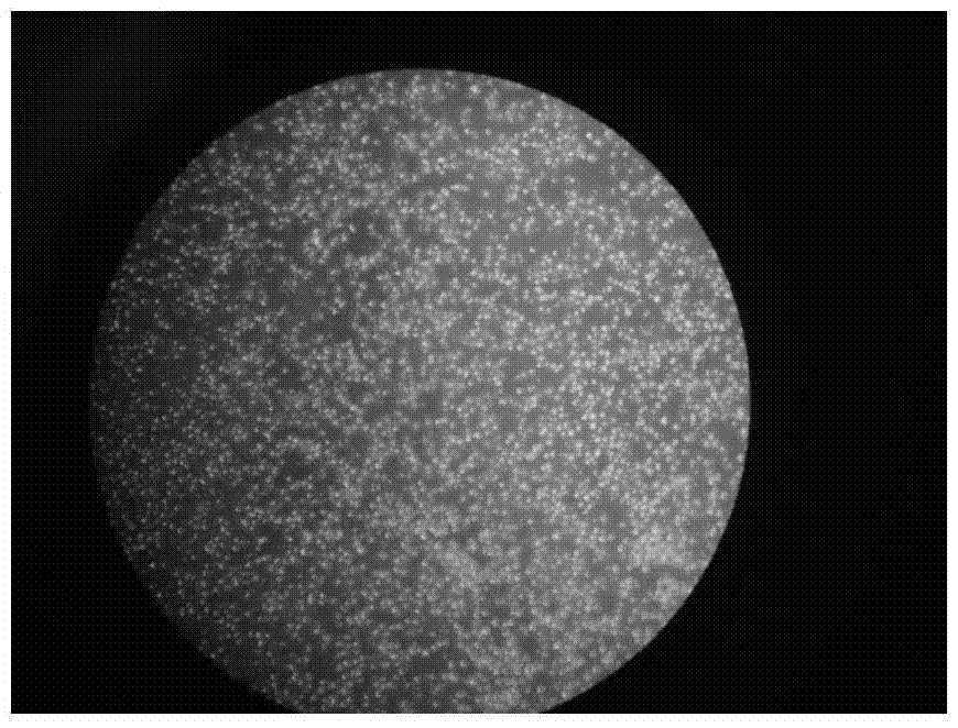
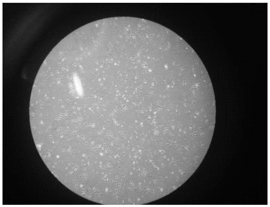
  • In-vitro culture method of vaginal epithelial cells of mouse
  • In-vitro culture method of vaginal epithelial cells of mouse
  • In-vitro culture method of vaginal epithelial cells of mouse

Examples


Embodiment 1

[0046] A method for culturing mouse vaginal epithelial cells in vitro, comprising the steps of:

[0047] Step (1), take the isolated vaginal tissue of the mouse, cut it into a size of 0.9cm×0.6cm, and then wash it with PBS containing double antibodies at 4°C as the cleaning solution. After cleaning until the cleaning solution is clear, the vaginal tissue Mix with 2U / ml dispase II at a volume ratio of 1:1.9, and freeze overnight at 4°C;

[0048] Step (2), separate the vaginal tissue cultured overnight in step (1), separate the epithelial layer tissue and lamina propria tissue, discard the lamina propria tissue, and then mix the epithelial layer tissue with 0.25% trypsin-0.02% EDTA according to 1 : Mixed at a volume ratio of 1.9, digested the vaginal epithelial tissue at 37°C for 9.5 minutes, then blown the epithelial layer with a sterile gun tip until the digested vaginal epithelial tissue cells were blown away, and then digested the vaginal epithelial tissue at 37°C for 9.5 mi...

Embodiment 2

[0051] A method for culturing mouse vaginal epithelial cells in vitro, comprising the steps of:

[0052] Step (1), take the isolated vaginal tissue of the mouse, cut it into a size of 1.1cm×0.4cm, and then wash it with PBS containing double antibody at 4°C as the cleaning solution. After cleaning until the cleaning solution is clear, the vaginal tissue Mix with 2U / ml dispase type II at a volume ratio of 1:2.1, and freeze overnight at 4°C;

[0053] Step (2), separate the vaginal tissue cultured overnight in step (1), separate the epithelial layer tissue and lamina propria tissue, discard the lamina propria tissue, and then mix the epithelial layer tissue with 0.25% trypsin-0.02% EDTA according to 1 : Mix at a volume ratio of 2.1, digest the vaginal epithelial tissue at 37°C for 10.5 minutes, then blow the epithelial layer with a sterile gun tip until the digested vaginal epithelial tissue cells are blown away, and then digest the vaginal epithelial tissue at 37°C for 10.5 minut...

Embodiment 3

[0056] A method for culturing mouse vaginal epithelial cells in vitro, comprising the steps of:

[0057] Step (1), take the isolated vaginal tissue of a mouse, cut it into 1cm×0.5cm size and place it in a 6-well plate, and then wash it with PBS containing double antibody at 4°C until the last time The washed PBS is not turbid, for a total of 10 minutes, transfer the vaginal tissue to a new 6-well plate, add 1ml 2U / ml Dispase II, 2ml 4°C refrigerator overnight;

[0058] Step (2): Separate the vaginal tissue cultured overnight in step (1) with ophthalmic tweezers on the ultra-clean bench, separate the epithelial tissue and lamina propria tissue, discard the lamina propria tissue, and add 0.25 %pancreatin-0.02% EDTA1ml, digest the vaginal epithelial tissue at 37°C for 10 minutes, then gently blow and beat the epithelial layer with a sterile probe for 30 seconds, then digest the vaginal epithelial tissue at 37°C for 10 minutes, and then gently blow and beat the epithelial layer fo...


PUM


Abstract

The invention relates to an in-vitro culture method of vaginal epithelial cells of a mouse and belongs to the technical field of cell culture. According to the method, isolated vaginal tissue of the mouse is sheared into pieces in the size of 1 cm*0.5 cm, washed and subjected to overnight culture through Dispase II, the vaginal tissue subjected to overnight culture is separated, lamina propria tissue is discarded, epithelial layer tissue is mixed with 0.25% pancreatin and 0.02% EDTA (ethylene diamine tetraacetic acid) and digested, finally, digestion is stopped through DMEM (Dulbecco modified Eagle medium) / F12 containing 10% fetal calf serum, obtained cell suspension is filtered, centrifugalized, re-suspended with a PBS (phosphate buffer solution) and EPILIFE sequentially and put into a 5% CO2 culture box with the temperature of 37 DEG C to be cultured, and a solution is replaced for the first time after two days and is replaced once every two days later. The method is convenient to operate, a large quantity of cells are obtained, the purity is high, the activity is high, the cells can be subcultured for 4-5 generations stably, and seed cells can be provided for observation of physiological and pathological change and the cancerization process as well as establishment of tissue-engineered vaginas of vaginal epithelium.

Description

technical field [0001] The invention belongs to the technical field of cell culture, and in particular relates to a method for culturing mouse vaginal epithelial cells in vitro, in particular to a method for primary culture of mouse vaginal epithelium by adopting an enzymatic digestion method. Background technique [0002] For the in vitro culture of mouse vaginal epithelial cells, domestic and foreign studies have used the tissue block method to culture vaginal epithelial cells. Although the operation is simple, it takes a long time and the passage cycle is long. [0003] In recent years, the mouse vaginal epithelium was isolated by enzymatic digestion, and the epithelial layer was digested into a single cell suspension by enzymatic digestion. After successful culture, it was used for tissue engineering vaginal research, but due to the However, due to the small number of cells obtained, the inability to adhere to the wall, and the poor ability of passage, there has not been...

Claims


Application Information

Patent Timeline
IPC IPC(8): C12N5/071
CPCC12N5/0682C12N2509/00
Inventor 祁文瑾刘燕燕
Owner 祁文瑾
Learn More