A preparation method of 3D printing bone orthopedic brace based on medical images

A medical imaging and orthopedic brace technology, applied in the field of medical devices, can solve the problems of unable to provide night brace information, difficult to obtain patient body surface data, difficult to guarantee the orthopedic effect, etc. easy effect

Inactive Publication Date: 2017-09-12
GUANGDONG KENVOXEL MEDICAL TECH CO LTD
View PDF8 Cites 1 Cited by
  • Summary
  • Abstract
  • Description
  • Claims
  • Application Information

AI Technical Summary

Problems solved by technology

[0006] Traditional orthopedic braces have the following problems: the stereotyped orthopedic effect of the orthopedic brace is limited by the professional level of the brace technician, and more depends on the experience of the brace technician, so the orthopedic effect is difficult to guarantee; the brace is too large and it is not easy to cover; The brace is not breathable, and it is very hot to wear in summer
Although the existing technology solves the problem of wearing comfort of the brace, it only relies on X-rays to obtain bone information, and the available information is limited
Because most of the X-ray films are positive and lateral line films in the standing state, it is impossible to provide information on night braces
In addition, most hospitals are not equipped with 3D scanners, making it difficult to obtain patient body surface data

Method used


Image

  • A preparation method of 3D printing bone orthopedic brace based on medical images

Examples


Embodiment

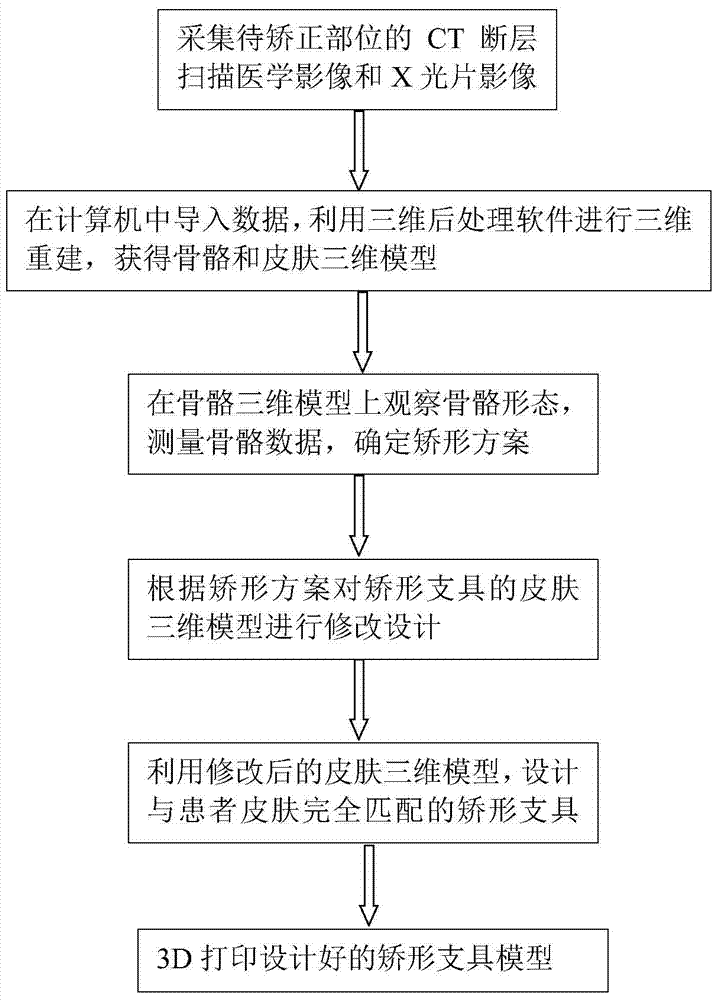
[0040] The present invention first collects medical images such as CT and MRI of the patient, reconstructs the three-dimensional model of the bone and the three-dimensional model of the skin of the part to be corrected in the computer, and designs a brace that fits the surface of the part to be corrected in a forward direction according to the three-dimensional model of the skin. The three-dimensional model determines the force point required for orthopedics and designs the brace to obtain a three-dimensional model of the orthopedic brace, which is finally printed and produced by a 3D printer. see figure 1 , including the following steps:

[0041] Step 1: Collect CT tomographic medical images and X-ray images of the parts to be corrected; obtain DICOM data.

[0042] Step 2: Import 2D medical imaging DICOM data into the computer, use 3D post-processing software to perform 3D reconstruction, and separate the bone 3D model and skin 3D model according to the density of different ...


PUM


Abstract

The invention provides a 3D printing skeleton correcting brace manufacturing method based on medical images. At first, the medical images such as CT and MRI of a patient are collected, a skeleton three-dimensional model and a skin three-dimensional model of a part to be corrected are reconstructed in a computer, a brace completely attached to the surface of the part to be corrected is designed forward according to the skin three-dimensional model, a force application point needed by correction is determined according to the skeleton three-dimensional model, the brace is designed, and a three-dimensional model of the correcting brace is obtained, and printed and produced by means of a 3D printer. The medical images such as the CT and MRI of the patient are utilized for carrying out three-dimensional reconstruction, skeleton deformity can be displayed well, and skeleton data are measured conveniently; meanwhile, the constructed three-dimensional model is utilized for directly extracting the skin model of the patient to serve as body surface data, the process for obtaining the body surface data of the patient is simplified, and the data are more accurate. The method is particularly suitable for preparing the correcting brace worn at night.

Description

technical field [0001] The invention relates to the field of medical devices, in particular to a 3D printing preparation method for bone orthopedic braces. Background technique [0002] Scoliosis is a three-dimensional deformity of the spine that includes an abnormal sequence of vertebral bodies in the coronal, sagittal, and horizontal planes. The spine of a normal person should be straight when viewed from the back, and the sides of the trunk should be symmetrical. If the shoulders are not equal in height from the front or the back is uneven from the back, "scoliosis" should be suspected. At this time, a standing X-ray of the entire spine is taken. If the frontal X-ray shows a lateral curvature of the spine greater than 10 degrees, it can be diagnosed as scoliosis. Mild scoliosis usually causes no obvious discomfort, and no obvious physical deformity can be seen in appearance. Severe scoliosis will affect the growth and development of infants and adolescents, deforming t...

Claims


Application Information

Patent Timeline
Patent Type & Authority Patents(China)
IPC IPC(8): A61F5/01A61B6/03A61B5/055
Inventor 庄永锋杨贻民
Owner GUANGDONG KENVOXEL MEDICAL TECH CO LTD
Why Eureka
Try Eureka