CT image denoising method based on principal component analysis

A principal component analysis and CT image technology, applied in the medical field, can solve the problem of not being able to better preserve image edges and details

Inactive Publication Date: 2015-11-11
嘉恒医疗科技(上海)有限公司
View PDF3 Cites 0 Cited by
  • Summary
  • Abstract
  • Description
  • Claims
  • Application Information

AI Technical Summary

Problems solved by technology

However, the main problem of the current common CT image denoising method is that it cannot preserve the edge and detail information of the image while filtering out the noise.

Method used


Image

  • CT image denoising method based on principal component analysis
  • CT image denoising method based on principal component analysis
  • CT image denoising method based on principal component analysis

Examples


Embodiment Construction

[0022] The present invention will be described in further detail below in conjunction with the accompanying drawings and specific embodiments, but not as a limitation of the present invention.

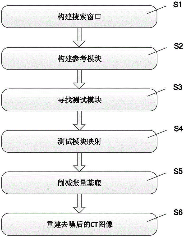
[0023] figure 1 It is a flow chart of the CT image denoising method based on principal component analysis of the present invention, comprising the following steps:

[0024] Step S1,

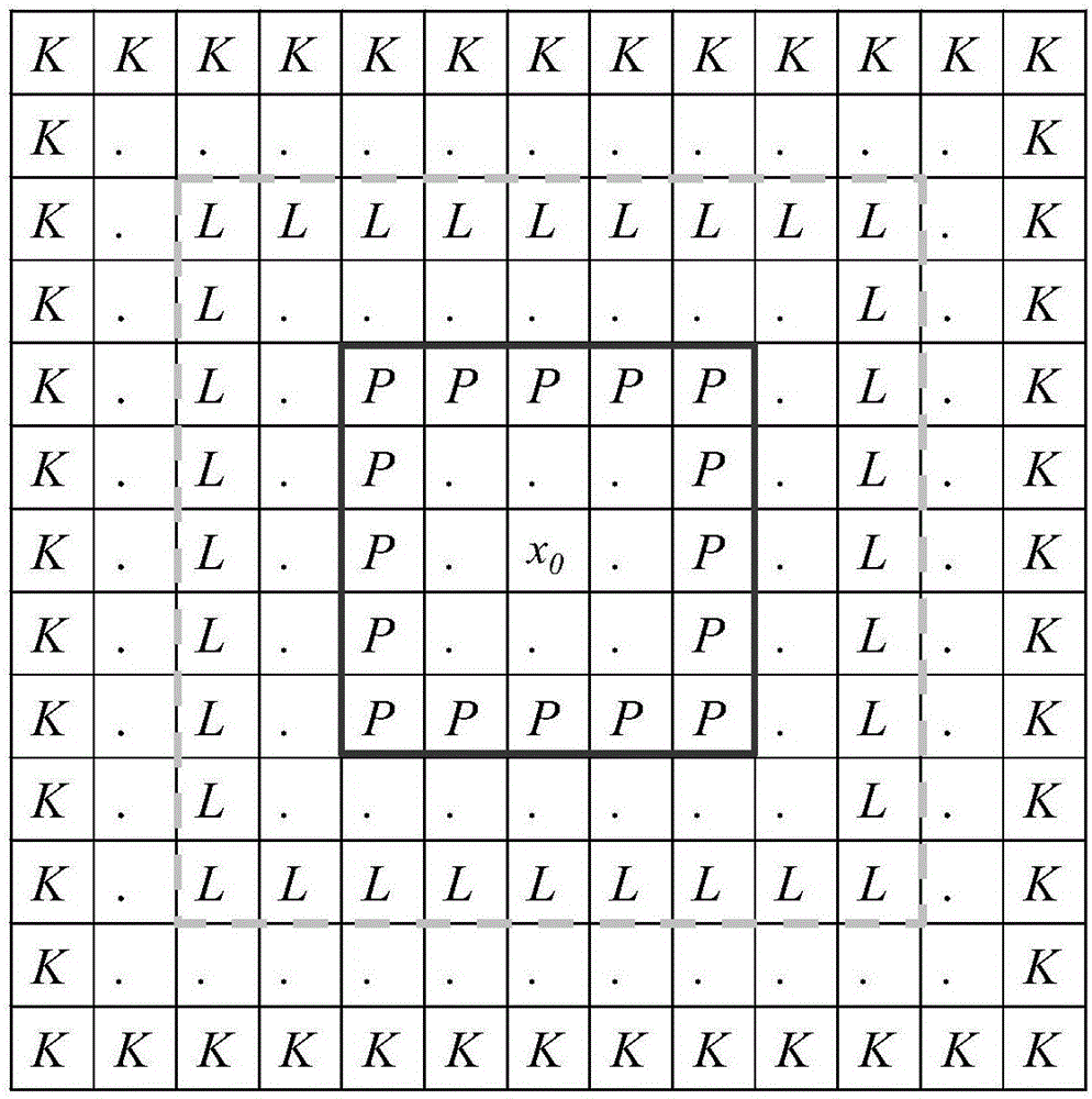
[0025] Build an adaptively resizable search window.

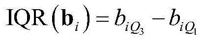
[0026] The search window can be a rectangular window, and the size of the search window can be automatically adjusted according to the characteristics of different windows. For example, construct a search window of size L×L, introduce two evaluation criteria to describe the weight of search windows of different sizes: median absolute deviation and interquartile range absolute deviation, define two windows K×K and L× L, and K>L, the optimal search window B can be obtained by comparing the weights of all windows in the range [P+1,K]. Expand all compa...


PUM


Abstract

The invention discloses a CT image denoising method based on principal component analysis. The method comprises steps: a search window is constructed, and the size of the search window can be adjusted adaptively; With a pixel containing noise in a CT image as a center, a rectangular reference module is constructed; in the search window, test modules similar to the reference module are searched, and a test module set is constructed; the test module set is mapped to a tensor substrate space of the principal component of the CT image; the substrate of tensor containing noise is subjected to reduction; the test modules are mapped to image space, and a CT image after denoising is reconstructed. Through the method, noise in a CT image can be removed accurately, and edge and detail information in the CT image can be reserved.

Description

technical field [0001] The invention relates to the field of medical technology, in particular to a CT image denoising method based on principal component analysis. Background technique [0002] Biomedical image processing is an important aspect of digital image processing. A lot of biomedical information is expressed in the form of images, such as CT images, which enable human vision to extend from the surface to the inside, and people can use them to obtain useful information on the anatomical shape, biochemical and physiological functions of the internal organs of the human body. Because the lesions in CT images are similar in grayscale and shape, it is difficult to distinguish them with the naked eye. People with different abilities and backgrounds often get different results on the same medical image. It is even more impossible to have a quantitative evaluation of the relative images. Therefore, the primary task of using a computer to post-process CT images is to enh...

Claims


Application Information

Patent Timeline
Patent Type & Authority Applications(China)
IPC IPC(8): G06T5/00
Inventor 郑重吴文波杨文晖赖暖翔
Owner 嘉恒医疗科技(上海)有限公司
Why Eureka
Try Eureka