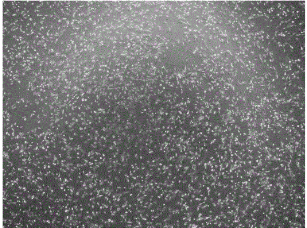
High-activity primary cartilage cell preparing method
A technology of chondrocytes and high vitality, applied in the direction of bone/connective tissue cells, animal cells, vertebrate cells, etc., can solve the problems of primary chondrocytes falling off and low yield, shorten the time of digestion and improve the yield rate, damage avoidance
- Summary
- Abstract
- Description
- Claims
- Application Information
AI Technical Summary
Problems solved by technology
Method used
Image
Examples
Embodiment 1
[0029] 1. At 24 hours after birth, 6 male or female mice are not limited to 6 young mice. Take the cartilage from the joints of the limbs and try to remove the clean soft tissue. The remaining transparent cartilage tissue is placed in a petri dish filled with PBS buffer and washed several times until there is residual The blood is rinsed clean. Preparation of PBS buffer: PBS tablets were purchased from Biowest, and the PBS tablets were directly dissolved in water to obtain PBS buffer.
[0030] 2. Add a small amount (about 1ml) of 0.1% mass fraction of type II collagenase (C6885, Sigma-Aldrich) to keep the tissue moist during the cutting process. Since the cartilage tissue is elastic and comes from young mice, the tissue mass is relatively small. Ordinary ophthalmic scissors are easy to slip off when cut, and they are not easy to cut, which directly affects the degree of collagenase digestion and ultimately affects the yield of cartilage cells. In the present invention, a surgica...
Embodiment 2
[0034] 1. At 24 hours after birth, 6 male or female mice are not limited to 6 young mice. Take the cartilage from the joints of the limbs and try to remove the clean soft tissue. The remaining transparent cartilage tissue is placed in a petri dish filled with PBS buffer and washed several times until there is residual The blood is rinsed clean. Preparation of PBS buffer: PBS tablets were purchased from Biowest, and the PBS tablets were directly dissolved in water to obtain PBS buffer.
[0035] 2. Add a small amount (about 1ml) of 0.1% mass fraction of type II collagenase (C6885, Sigma-Aldrich) to keep the tissue moist during the cutting process. Since the cartilage tissue is elastic and comes from young mice, the tissue mass is relatively small. Ordinary ophthalmic scissors are easy to slip off when cut, and they are not easy to cut, which directly affects the degree of collagenase digestion and ultimately affects the yield of cartilage cells. In the present invention, a surgica...
Embodiment 3
[0039] 1. At 24 hours after birth, 6 male or female mice are not limited to 6 young mice. Take the cartilage from the joints of the limbs and try to remove the clean soft tissue. The remaining transparent cartilage tissue is placed in a petri dish filled with PBS buffer and washed several times until there is residual The blood is rinsed clean. Preparation of PBS buffer: PBS tablets were purchased from Biowest, and the PBS tablets were directly dissolved in water to obtain PBS buffer.
[0040] 2. Add a small amount (about 1ml) of 0.1% mass fraction of type II collagenase (C6885, Sigma-Aldrich) to keep the tissue moist during the cutting process. Since the cartilage tissue is elastic and comes from young mice, the tissue mass is relatively small. Ordinary ophthalmic scissors are easy to slip off when cut, and they are not easy to cut, which directly affects the degree of collagenase digestion and ultimately affects the yield of cartilage cells. In the present invention, a surgica...
PUM
 Abstract
Description
Claims
Application Information
 - R&D
- Intellectual Property
- Life Sciences
- Materials
- Tech Scout
- Unparalleled Data Quality
- Higher Quality Content
- 60% Fewer Hallucinations
Browse by: Latest US Patents, China's latest patents, Technical Efficacy Thesaurus, Application Domain, Technology Topic, Popular Technical Reports.
© 2025 PatSnap. All rights reserved.Legal|Privacy policy|Modern Slavery Act Transparency Statement|Sitemap|About US| Contact US: help@patsnap.com


