Eureka AIR delivers breakthrough ideas for toughest innovation challenges, trusted by R&D personnel around the world.

A minimally invasive intramedullary fixation device for pelvic fractures

A technology of pelvic fractures and fixation devices, applied in the direction of internal fixators, fixators, internal bone synthesis, etc., to achieve the effect of firm fixation, pain relief, and light damage

Inactive Publication Date: 2017-03-01
陈卫 +1
View PDF7 Cites 5 Cited by
  • Summary
  • Abstract
  • Description
  • Claims
  • Application Information

AI Technical Summary

Problems solved by technology

With the development of minimally invasive technology, the use of minimally invasive surgery in orthopedic surgery is becoming more and more common, but there is no precedent for the use of intramedullary fixation in the surgical treatment of pelvic ring and acetabular fractures, especially the use of reconstruction plates for reduction and fixation It is difficult to be satisfied with the effect, so intramedullary fixation in pelvic ring and acetabular fracture surgery has become a difficult problem to solve at present

Method used


Image

  • A minimally invasive intramedullary fixation device for pelvic fractures
  • A minimally invasive intramedullary fixation device for pelvic fractures
  • A minimally invasive intramedullary fixation device for pelvic fractures

Examples


Embodiment Construction

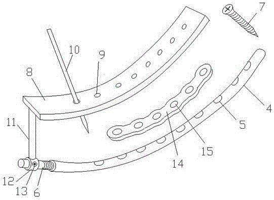
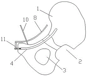
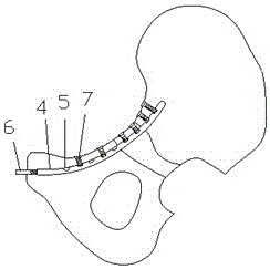
[0020] The present invention includes a guide wire, a guide sleeve, a soft drill, an arc-shaped intramedullary nail 4, an arc-shaped screw guide plate 8, a guide pin 10, a fixing screw 7, an intramedullary nail tail cap 6, a connecting rod 11, and a reconstruction plate 14.

[0021] The guide wire and the guide sleeve of the present invention are curved arcs, the arc matches the radian of the preset pelvic intramedullary tunnel, the outer diameter of the guide sleeve matches the preset inner diameter of the pelvic intramedullary tunnel, and is soft. The drill is located in the guide sleeve, and the guide wire is threaded on the central axis of the soft drill. During the operation, firstly, the soft drill drills the pelvic intramedullary tunnel on the pelvis according to the preset pelvic intramedullary tunnel path under the guidance of the guide wire and the guide sleeve, and then puts the arc-shaped intramedullary nail 4 in the tunnel, Immobilize the fracture site.

[0022] ...


PUM


Abstract

A minimally invasive intramedullary fixation device for pelvic fractures belongs to the technical field of orthopedic medical devices and is used for intramedullary reduction and fixation of pelvic fractures. The technical plan is: design the pelvic intramedullary tunnel in advance under the three-dimensional reconstruction of pelvic CT, make the guide wire and guide sleeve according to the curvature of the pelvic intramedullary tunnel, and use a soft drill to drill under the guidance of the pre-bent guide wire and C-arm fluoroscopy. Out of the pelvic intramedullary tunnel, put the arc-shaped intramedullary nail into the pelvic intramedullary tunnel along the guide wire, and fix the screws to securely connect the arc-shaped intramedullary nail to the pelvis. intramedullary nail. The pelvic intramedullary fixation adopted in the present invention is an innovation in the pelvic fracture fixation operation. This intramedullary fixation device and method can realize the perfect reduction and fixation of the pelvic ring and acetabular fractures with the cooperation of direct vision and C-arm irradiation. It is convenient and firmly fixed, which greatly shortens the operation time and recovery time, improves the treatment effect, and reduces the pain of the patient.

Description

technical field [0001] The invention relates to an intramedullary fixation device for open reduction and fixation of pelvic fractures, belonging to the technical field of orthopedic medical devices. Background technique [0002] Pelvic internal fixation is a new technology in the field of orthopedic trauma treatment in recent years, and it also shows a new direction for the development of orthopedic surgery medical technology. Compared with traditional surgery, intramedullary bone treatment of the pelvis has the advantages of better support and less damage to surrounding tissues, and is more and more popular among doctors and patients. The development of contemporary science and technology provides a strong guarantee for intramedullary fixation. Under the guidance of X-ray machines, CT and advanced electronic and optical equipment, doctors can use special instruments and various instruments to perform operations. With the development and progress of science and technology, ...

Claims


Application Information

Patent Timeline
Patent Type & Authority Patents(China)
IPC IPC(8): A61B17/72A61B17/80
CPCA61B17/00234A61B17/725A61B17/8066A61B2017/564
Inventor 杜晨光陈伟侯志勇张英泽吕红芝魏宁常文利
Owner 陈卫
Why Eureka
Eureka Blog
Learn More