


Quick Research
Generate reliable direction feasibility study reports for your R&D in just a few steps.

Technical Q&A
Discover and master advanced knowledge NOW. Basics, ideas, possibilities, all at once.

Find Solutions
As an expert in R&D theories, this can generate solutions to your technical problems instantly.

Evaluate Feasibility
Analyze your overall solution with one click, know your potential R&D risks in advance.

Monitor Landscape
Get weekly tech updates, stay abreast of the latest tech innovations and key insights.
Image processing device, image processing method, and image processing program
An image processing device and image technology, applied in image data processing, image data processing, image enhancement and other directions, can solve the problems of inability to calculate feature quantities, inability to discriminate between normal and abnormal with high precision, and achieve the effect of high-precision recognition
- Summary
- Abstract
- Description
- Claims
- Application Information
AI Technical Summary
Problems solved by technology
Method used
Image

Examples
Embodiment approach 1
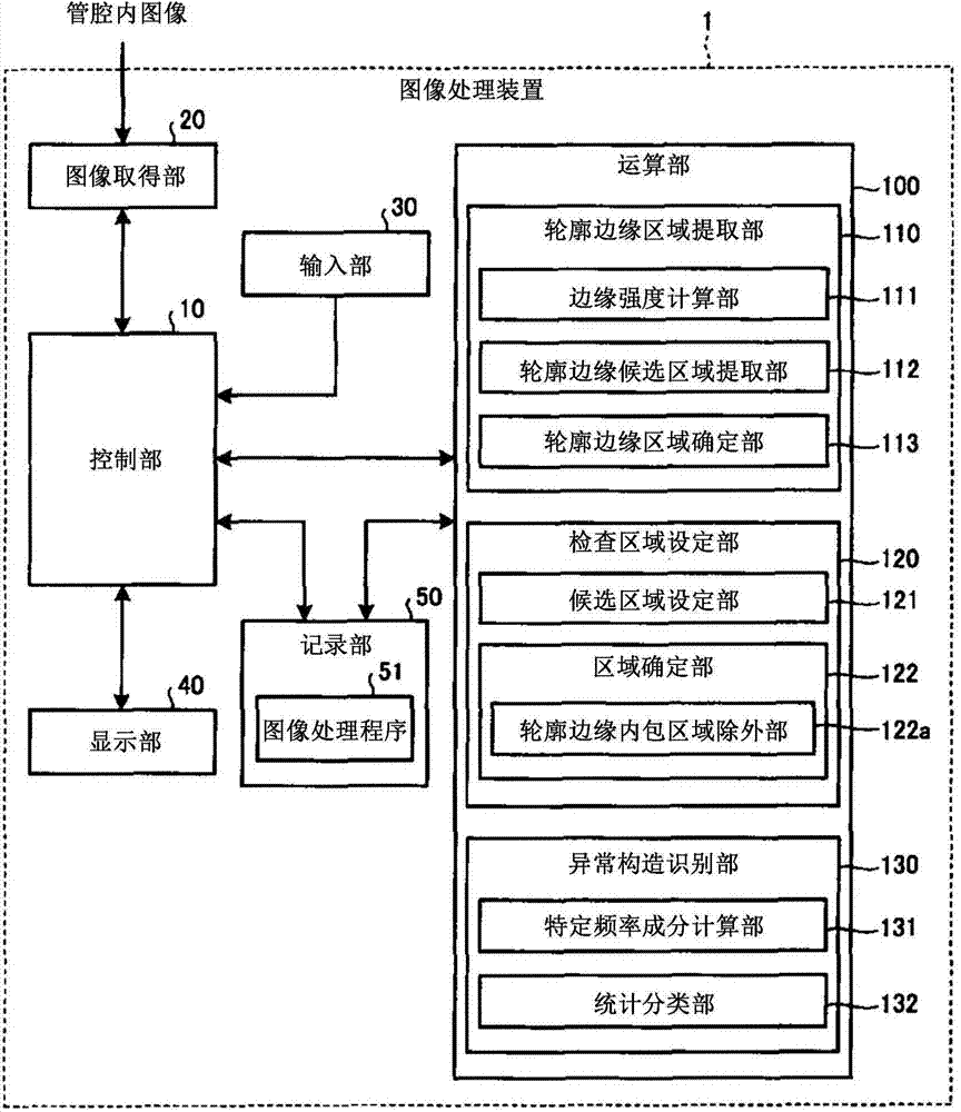
[0051] figure 1 It is a block diagram showing the image processing device according to Embodiment 1 of the present invention. like figure 1 As shown, the image processing device 1 has a control unit 10 that controls the overall operation of the image processing device 1, an image acquisition unit 20 that acquires image data corresponding to an image captured by an endoscope, and accepts input from the outside. An input unit 30 for input signals, a display unit 40 for performing various displays, a recording unit 50 for storing image data acquired by the image acquisition unit 20 and various programs, and a computing unit 100 for performing predetermined image processing on the image data.
[0052] The control unit 10 is realized by hardware such as a CPU, reads various programs recorded in the recording unit 50, and executes the configuration of the image processing device 1 based on the image data input from the image acquisition unit 20 and the operation signal input from t...
Deformed example 1-1
[0091] Next, Modification 1-1 of Embodiment 1 of the present invention will be described.
[0092] In Embodiment 1 above, an example of using the spatial frequency component as the texture information of the mucous membrane surface was shown, but instead of the spatial frequency component, a statistical feature value using a co-occurrence matrix or a Local Binary Pattern (local binary pattern) may be used. , high-order local autocorrelation, SIFT (Scale-Invariant Feature Transform: scale-invariant feature transformation), HOG (Histograms of Oriented Gradients: gradient direction histogram) and other well-known texture information.
Deformed example 1-2
[0094] Next, Modification 1-2 of Embodiment 1 of the present invention will be described.
[0095] Figure 9 It is a block diagram showing the configuration of the computing unit included in the image processing device of Modification 1-2. Figure 9 The illustrated calculation unit 100A includes a contour edge region extraction unit 110A, an inspection region setting unit 120A, and an abnormal structure recognition unit 130A. In addition, the configuration and operation of the image processing device other than the computing unit 100A are the same as those of Embodiment 1 (see figure 1 ).
[0096] Contour edge region extracting unit 110A is relative to figure 1 The shown contour edge region extraction section 110 also has a low-absorption wavelength selection section 114 . The low-absorption wavelength selection unit 114 selects a wavelength component (low-absorption wavelength component) in which the degree of absorption or scattering in the living body is the lowest amon...
PUM
 Abstract
Description
Claims
Application Information
 - R&D Engineer
- R&D Manager
- IP Professional
- Industry Leading Data Capabilities
- Powerful AI technology
- Patent DNA Extraction
Browse by: Latest US Patents, China's latest patents, Technical Efficacy Thesaurus, Application Domain, Technology Topic, Popular Technical Reports.
© 2024 PatSnap. All rights reserved.Legal|Privacy policy|Modern Slavery Act Transparency Statement|Sitemap|About US| Contact US: help@patsnap.com