
X-ray imaging equipment

An imaging device and X-ray technology, applied in the field of nuclear imaging, can solve problems such as unfavorable carrying, increased radiation, and increased damage to human health, and achieve the effects of increasing portability, reducing volume, and reducing damage

Inactive Publication Date: 2015-03-25
SHENZHEN INST OF ADVANCED TECH
View PDF8 Cites 6 Cited by
  • Summary
  • Abstract
  • Description
  • Claims
  • Application Information

AI Technical Summary

Problems solved by technology

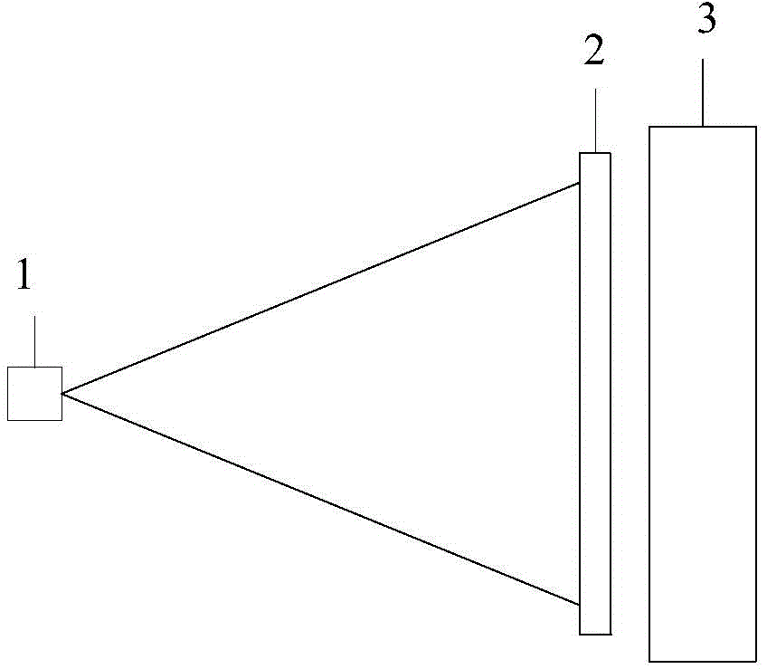
The structure of ordinary X-ray DR is as follows figure 1 As shown, it includes a point X light source 1, an irradiated object 2 and a flat panel detector 3, which has the following disadvantages: (1) Using a point X light source 1 makes hand bone images easy to overlap, thus affecting the doctor's assessment of bone age. (2) In order to avoid image overlap, the distance between the irradiated object 2 and the point X light source 1 is increased, resulting in a larger volume of the entire photographic equipment, which is not conducive to carrying; it increases the radiation dose and increases unnecessary radiation, which increases the damage to human health, especially for children

Method used


Image

  • X-ray imaging equipment
  • X-ray imaging equipment
  • X-ray imaging equipment

Examples


Embodiment Construction

[0030] In order to make the object, technical solution and advantages of the present invention clearer, the present invention will be described in further detail below in conjunction with the embodiments and accompanying drawings. Here, the exemplary embodiments and descriptions of the present invention are used to explain the present invention, but not to limit the present invention.

[0031] The inventors found that because the common X-ray DR uses a point X light source 1, it has the following disadvantages: (1) Using a point X light source 1 makes hand bone images easy to overlap, thereby affecting the doctor's determination of bone age; 2) In order to avoid image overlap, the distance between the irradiated object 2 and the point X light source 1 must be increased, resulting in a large volume of the entire photographic equipment, which is not conducive to carrying; the radiation dose increases and unnecessary radiation is increased , thus increasing the damage to human he...


PUM


Abstract

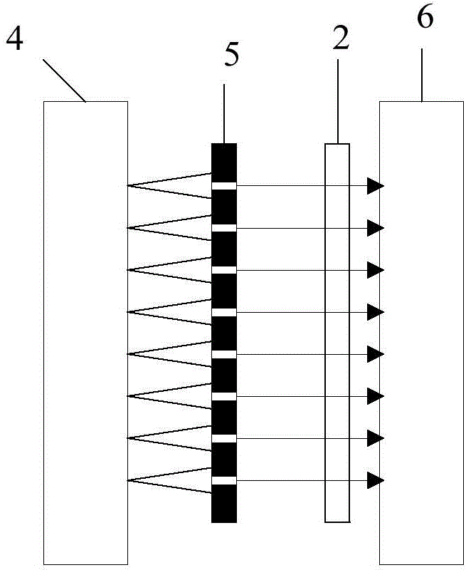
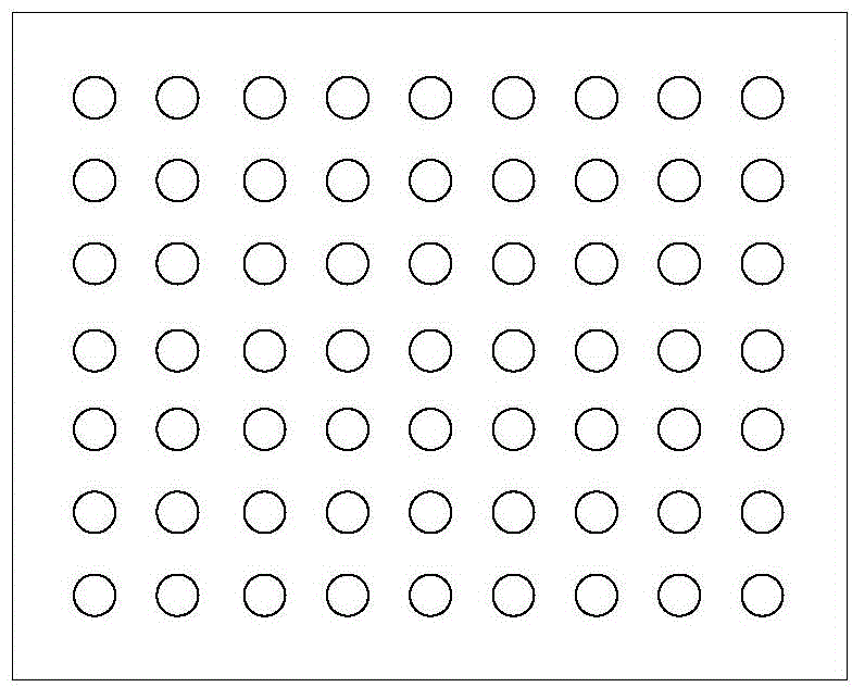
The invention provides X-ray imaging equipment. The X-ray imaging equipment comprises an array X-ray source, a collimator and an X-ray flat panel detector, wherein the array X-ray source is used for emitting array X-ray beams; the collimator is used for collimating the emitted array X-ray beams and irradiating the collimated array X-ray beams to a test object; the X-ray flat panel detector is used for receiving the array X-ray beams penetrating through the test object, and an X-ray photography image is formed by utilization of the received array X-ray beams. By the adoption of the combination of the array X-ray source and the collimator, the X-ray beams approximating parallel and an area source is formed, and it is avoided that as hand bone images are easily overlapped, judgment of a doctor on the bone age is affected; compared with an ordinary X-ray DR technology, as the distance between the hand and the X source is not increased, the size of the whole equipment is reduced, and portability is improved; unnecessary radiation is reduced, and therefore injury to human health is reduced.

Description

technical field [0001] The invention relates to the technical field of nuclear imaging, in particular to an X-ray imaging device. Background technique [0002] The changes in the development of human bones are basically similar, and the development process of each bone has continuity and stages. Bones at different stages have different morphological characteristics, therefore, bone age assessment can more accurately reflect the level of growth and maturity of an individual. It can not only determine the biological age of children, but also can understand the growth and development potential of children and the trend of sexual maturity early through bone age: the adult height of children can also be predicted through bone age, and the determination of bone age is also helpful for the diagnosis of some pediatric endocrine diseases. Great help. Bone age was first used in medical research and to measure the growth and development of children, and then it was widely used to det...

Claims


Application Information

Patent Timeline
IPC IPC(8): A61B6/03
Inventor 陈垚郑海荣梁栋洪序达桂建保胡战利
Owner SHENZHEN INST OF ADVANCED TECH
Learn More