Imaging method and system applied to neurosurgery

A technology of neurosurgery and imaging method, applied in the field of neurosurgery imaging method and its system, can solve the problem of not being able to clearly and intuitively reflect the distribution of white matter fiber bundles

Active Publication Date: 2013-12-25
SHENZHEN INST OF ADVANCED TECH
View PDF4 Cites 9 Cited by
  • Summary
  • Abstract
  • Description
  • Claims
  • Application Information

AI Technical Summary

Problems solved by technology

However, the images obtained by applying DTI, fMRI and PMRI technologies alone still cannot clearly and intuitively reflect the distribution of each functional area of ​​the brain and the white matter fiber tracts connecting each functional area.

Method used


Image

  • Imaging method and system applied to neurosurgery
  • Imaging method and system applied to neurosurgery
  • Imaging method and system applied to neurosurgery

Examples


Embodiment Construction

[0035] In order to solve the problem that the images obtained by using DTI, fMRI and PMRI technologies alone cannot clearly and intuitively reflect the distribution of the functional areas of the brain and the white matter fiber bundles connecting the functional areas, this embodiment provides a neurosurgery Imaging method and system for surgery. The imaging method and system applied to neurosurgery will be specifically described below in conjunction with specific embodiments.

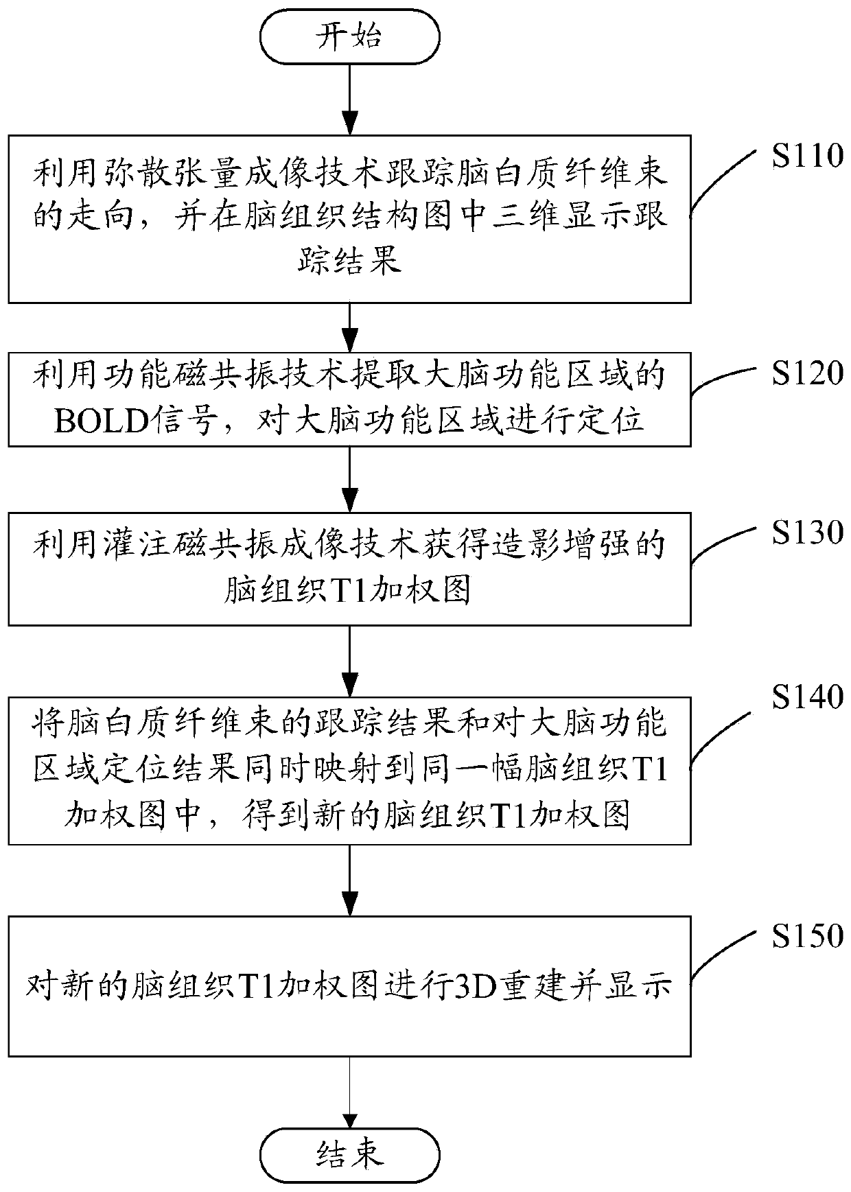
[0036] Please refer to figure 1 The imaging method applied to neurosurgery provided by this embodiment includes the following steps:

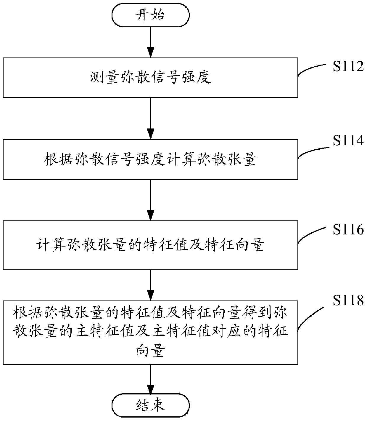
[0037] Step S110: using the diffusion tensor imaging technology to track the direction of the white matter fiber bundles, and display the tracking results in three dimensions in the brain histogram. Please refer to figure 2 , in this step, the steps of using diffusion tensor imaging technology to track the direction of white matter fiber bundles include:

[0038] S...


PUM


Abstract

The invention relates to an imaging method applied to a neurosurgery. The method includes the following steps: utilizing a diffusion tensor imaging technology to track the moving trend of brain white matter fiber bundles, displaying a tracking result in a three-dimensional mode in a brain tissue structure graph, utilizing a functional magnetic resonance technology to extract BOLD signals in brain functional regions to locate the brain functional regions, utilizing a filling magnetic resonance imaging technology to obtain a brain tissue T1 weighted graph obtained through contract enhancement, and mapping the tracking result of the brain white matter fiber bundles and a location result of the brain functional regions into the same brain tissue T1 weighted graph simultaneously to obtain a new brain tissue T1 weighted graph. By means of the imaging method applied to the neurosurgery, the tracking result of the brain white matter fiber bundles and the location result of the brain functional regions are mapped into the same brain tissue T1 weighted graph simultaneously, and distribution conditions of the brain functional regions and the white matter fiber bundles connecting the functional regions can be reflected clearly and visually. In addition, an imaging system applied to the neurosurgery is further provided.

Description

technical field [0001] The invention relates to the technical field of image processing, in particular to an imaging method and system applied to neurosurgery. Background technique [0002] Brain tumors have a high incidence rate and are fatal and disabling diseases that seriously endanger human health. Malignant brain tumors have the characteristics of "three highs and one low" in terms of incidence, recurrence rate, high mortality rate and low cure rate. For a long time, the effective means of targeting tumors is the first choice in the medical field to carry out effective and complete surgical resection of tumor tissues. In recent years, with the advancement of imaging diagnostic techniques and the application of microneurosurgery techniques, the diagnosis and treatment of brain tumors have made significant progress in general. However, no breakthrough has been made in the treatment of patients with malignant brain tumors (such as glioma, etc.). Because in tumor resecti...

Claims


Application Information

Patent Timeline
Patent Type & Authority Applications(China)
IPC IPC(8): A61B5/055
CPCA61B5/055G01R33/56341G01R33/56366G01R33/4806G01R33/5608A61B5/0263A61B5/1128A61B5/0036
Inventor 张娜刘新钟耀祖郑海荣
Owner SHENZHEN INST OF ADVANCED TECH
Why Eureka
Try Eureka