Automatic cell localization method based on minimized model L1

A cell positioning and minimization technology, applied in the field of biomedical optical image processing, can solve the problems that neuron cells cannot be processed well and are not mature enough

Inactive Publication Date: 2013-08-07
HUAZHONG UNIV OF SCI & TECH
View PDF4 Cites 8 Cited by
  • Summary
  • Abstract
  • Description
  • Claims
  • Application Information

AI Technical Summary

Problems solved by technology

However, this method does not work well for neuronal cells with thick neurites
The positioning of nerve cells still depends on human assistance, and the method of automatic positioning of nerve cells by computer is far from mature

Method used


Image

  • Automatic cell localization method based on minimized model L1
  • Automatic cell localization method based on minimized model L1
  • Automatic cell localization method based on minimized model L1

Examples


example

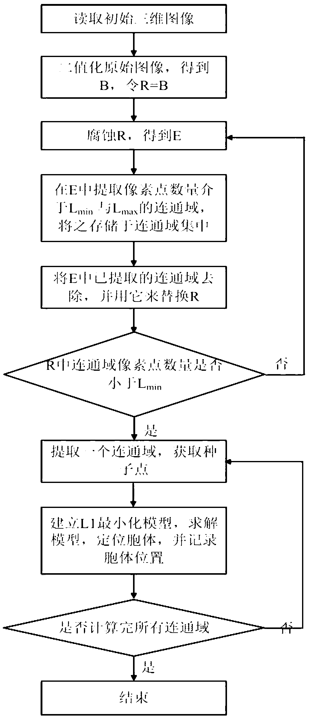
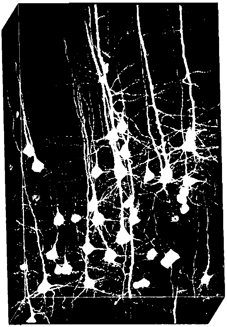
[0131] Taking mouse brain slice images acquired by super-resolution fluorescence imaging microscope or functional two-photon confocal imaging microscope as the object, the original images are preprocessed to facilitate subsequent operations.

[0132] step 1:

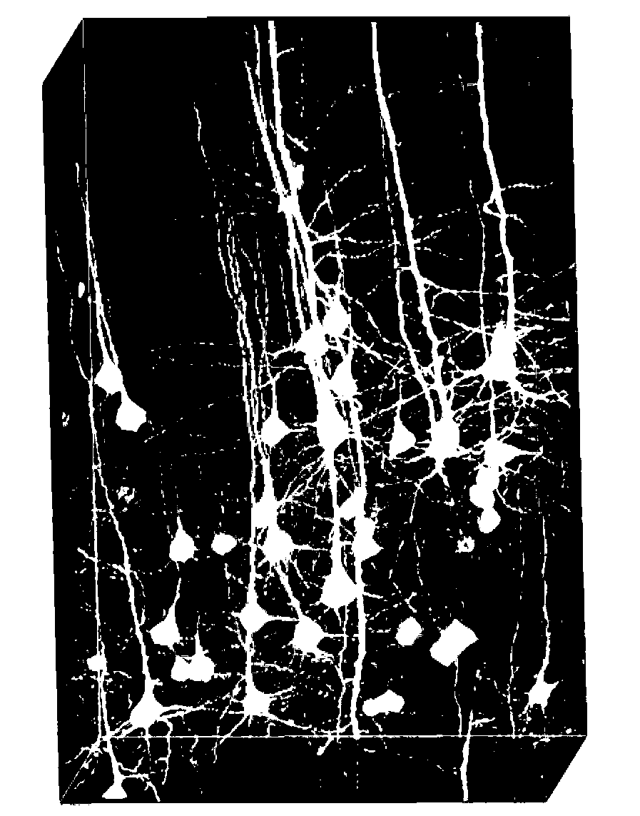
[0133] Read in the 3D original image (such as figure 2 shown), the four (each matt) pixels of each frame of the two-dimensional image of the three-dimensional original image are combined into one pixel, and the signal value of each pixel is directly added to obtain a new image Denoted as I (such as image 3 shown);

[0134] Make a small operation on I and T1 (T1 is preferably 400), and then perform 20 convolution operations with a 9x9x1 mean template, and the new image obtained is called the background image, which is recorded as C (such as Figure 4 shown);

[0135] According to formula I, utilize I and C to obtain binarized image, denote as B (such as Figure 5 shown);

[0136] Step 2:

[0137] According to the...


PUM


Abstract

The invention discloses an automatic cell localization method based on a minimized model L1. The automatic cell localization method includes (1) performing binarization processing to an original image, and acquiring a binarization image B; (2) etching the B, acquiring neuronal connected domains, and performing processing each connected domain according to the steps (3) and (4), and acquiring positions of all cells; (3) embedding each connected domain into a rectangle, namely a sub-region, acquiring candidate seed points from the sub-region, removing redundant candidate seed points according to spaces among the candidate seed points, taking rest candidate seed points as actual seed points, and marking the number of the actual seed points as k; and (4) establishing the minimized model L1, and acquiring actual cell positions and radius in the connected domains. By the aid of the automatic cell localization method, cell localization is performed in a sub-region extracting manner, calculation of each sub-region can be performed simultaneously and independently, all the connected domains can be calculated in a parallel manner, entire calculation speed can be greatly increased, so that wide-range data can be processed effectively.

Description

technical field [0001] The invention belongs to the field of biomedical optical image processing, in particular to a cell positioning method in biomedical optical images. Specifically, it is a fully automatic cell localization method based on an L1 minimization model, wherein L1 is a norm of 1. The method of the invention is particularly suitable for the localization of neuronal cell bodies. Background technique [0002] Neural circuits are the physical basis of brain function. Mapping fine-grained neural circuits could dramatically improve our understanding of brain function. If we do the work of locating nerve cells in advance, we will be able to trace neurites faster and more precisely, allowing us to efficiently map neural circuits. In fact, this method has been widely used in neuroscience. For example, we have successfully studied the dependence between cancer stem cells and the neural microenvironment, and found the distribution rules between nerve cells and nerve b...

Claims


Application Information

Patent Timeline
Patent Type & Authority Applications(China)
IPC IPC(8): G06T7/00
Inventor 曾绍群龚辉骆清铭李靖
Owner HUAZHONG UNIV OF SCI & TECH
Why Eureka
Try Eureka