
X-ray computed tomography scanning system and method

A tomography and computer technology, applied in the field of medical imaging, can solve the problem that the radiation dose cannot be reduced

Active Publication Date: 2015-04-08
SIEMENS SHANGHAI MEDICAL EQUIP LTD
View PDF6 Cites 0 Cited by
  • Summary
  • Abstract
  • Description
  • Claims
  • Application Information

AI Technical Summary

Problems solved by technology

[0004] It can be seen that according to the existing CT scanning technology, even if the three-dimensional ROI is only a small organ or tissue of the object to be examined, the total radiation dose cannot be reduced

Method used


Image

  • X-ray computed tomography scanning system and method
  • X-ray computed tomography scanning system and method
  • X-ray computed tomography scanning system and method

Examples


Embodiment Construction

[0047] In order to make the purpose, technical solution and advantages of the present invention clearer, the following examples are given to further describe the present invention in detail.

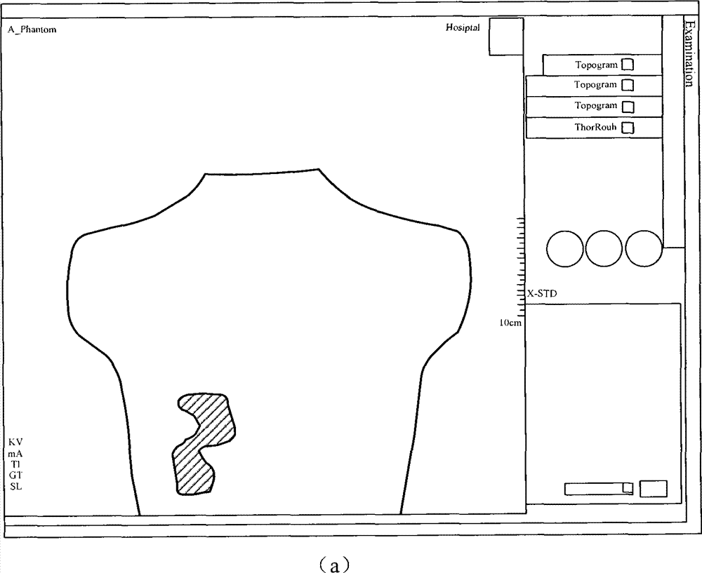
[0048] figure 1 It is a system structure diagram of an X-ray computed tomography system in a specific embodiment of the present invention.

[0049] Such as figure 1 As shown, in a specific embodiment of the present invention, the X-ray computed tomography system includes: a cross-sectional scanning parameter determination unit 10, which is used to determine the optimal radiation starting points required for scanning the regions of interest in each two-dimensional cross-section Angle α 0 opt and each minimum reconstruction angle γmin; the control curve determination unit 20 is used to obtain each optimal radiation start angle α 0 opt and each minimum reconstruction angle γmin, determine a control curve for controlling the scanning of the three-dimensional region of interest along the d...


PUM


Abstract

The invention relates to an X-ray computed tomography scanning system and a method. The X-ray computed tomography scanning system comprises a profile scanning parameter determining unit, a control curve determining unit and a scanner, wherein the profile scanning parameter determining unit is used for determining each optimal radiation starting angle alpha<0>opt and each minimum reconstruction angle gamma min which are required for scanning regions of interest in each two-dimensional profile; the control curve determining unit is used for determining control curves which are used for controlling scanning of three-dimensional regions of interest along the direction that a bedstead goes into or out of a gantry according to the obtained optimal radiation starting angles alpha<0>opt and the obtained minimum reconstruction angles gamma min; and the scanner is used for scanning the three-dimensional regions of interest according to the control curves. By means of the X-ray computed tomography scanning system and the method, image reconstruction of the three-dimensional regions of interest can be achieved by using few total radiation doses, energy consumption is saved, temporal resolution of image reconstruction is improved, and service life of X-ray tubes is prolonged.

Description

technical field [0001] The invention relates to the field of medical imaging, in particular to an X-ray computed tomography system and method. Background technique [0002] During X-ray computed tomography (Computed Tomography, CT), the organ or tissue to be inspected that requires high-quality image reconstruction is usually called a region of interest (ROI), and the ROI is a three-dimensional region. Based on the existing technology, no matter which part of the object to be scanned or the size of the ROI to be scanned and reconstructed is located, it must be determined according to the opening degree of the fixed collimator (phi-collimator), the fixed radiation dose and a certain scanning method The fixed reconstruction angle is used to perform radiological scans on the patient one by one. [0003] In the traditional full scan method, a full scan with a reconstruction angle (irradiation duration) γ of 360° is performed on each slice. In the partial scanning method, a fix...

Claims


Application Information

Patent Timeline
Patent Type & Authority Patents(China)
IPC IPC(8): A61B6/03
Inventor 张纪庄王薇
Owner SIEMENS SHANGHAI MEDICAL EQUIP LTD
Learn More