
Displacement detecting method and device thereof in elasticity imaging

A technology of displacement detection and elastic imaging, which is applied in measuring devices, organ movement/change detection, image analysis, etc., can solve the problems of poor image quality and large amount of calculation, and achieve the effect of reducing the amount of calculation and meeting the clinical real-time requirements

Active Publication Date: 2012-12-19
SHENZHEN MINDRAY BIO MEDICAL ELECTRONICS CO LTD
View PDF5 Cites 24 Cited by
  • Summary
  • Abstract
  • Description
  • Claims
  • Application Information

AI Technical Summary

Problems solved by technology

The image quality obtained by the method based on cross-correlation is good, but the amount of calculation is large, and there is a certain requirement for the echo data sampling rate; the method based on phase shift is fast to calculate, but there is a problem of phase aliasing, which is mainly suitable for the case of small displacement , and its lateral displacement is usually ignored, and the image quality is relatively poor; the method based on tissue Doppler is the easiest to implement, but it depends on the signal-to-noise ratio and processing method of the Doppler signal, and there are also The aliasing problem, and ignoring its lateral displacement, the image quality is relatively poor

Method used


Image

  • Displacement detecting method and device thereof in elasticity imaging
  • Displacement detecting method and device thereof in elasticity imaging
  • Displacement detecting method and device thereof in elasticity imaging

Examples


Embodiment Construction

[0024] The present invention will be further described in detail below through specific embodiments in conjunction with the accompanying drawings.

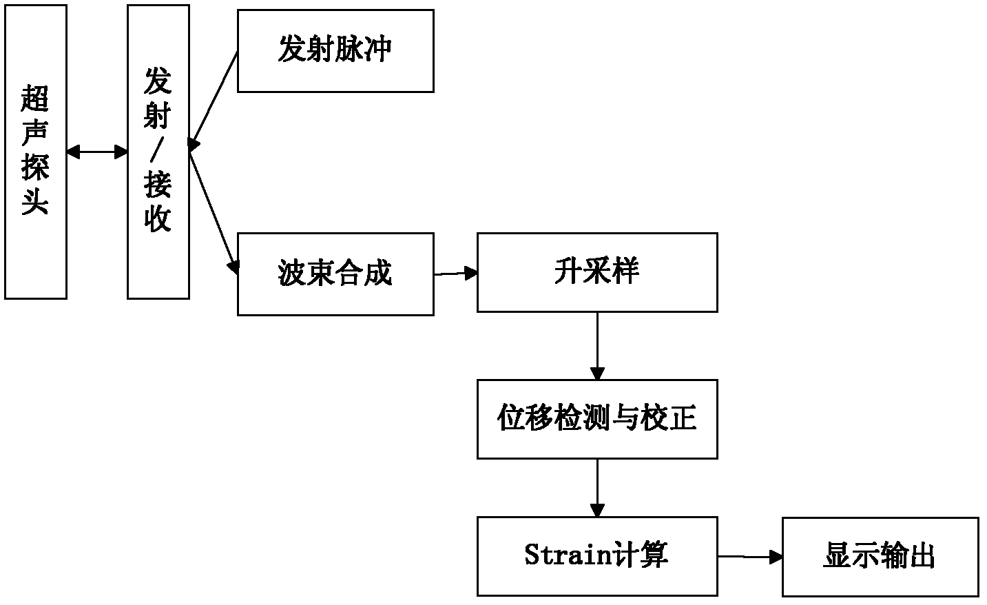
[0025] like figure 1 It is a schematic diagram of an embodiment of an elastography system of the present invention. In elastography mode, the ultrasonic probe transmits ultrasonic waves and receives echo information according to the pre-set scanning rules of the system, outputs radio frequency (radio frequency, RF) signals after the beamforming link, and then passes through the + into I / Q two-way baseband signals, and down-sampling process is performed at the same time, so that the sampling rate of I / Q signals is reduced. The down-sampling rate is preset by the system, and then through the guided phase zero estimation (GPZE, guided phase zero estimation) algorithm In the displacement detection link, a frame of displacement result is calculated by using a pair of I / Q signal frames each time, and then through the strain calculation...


PUM


Abstract

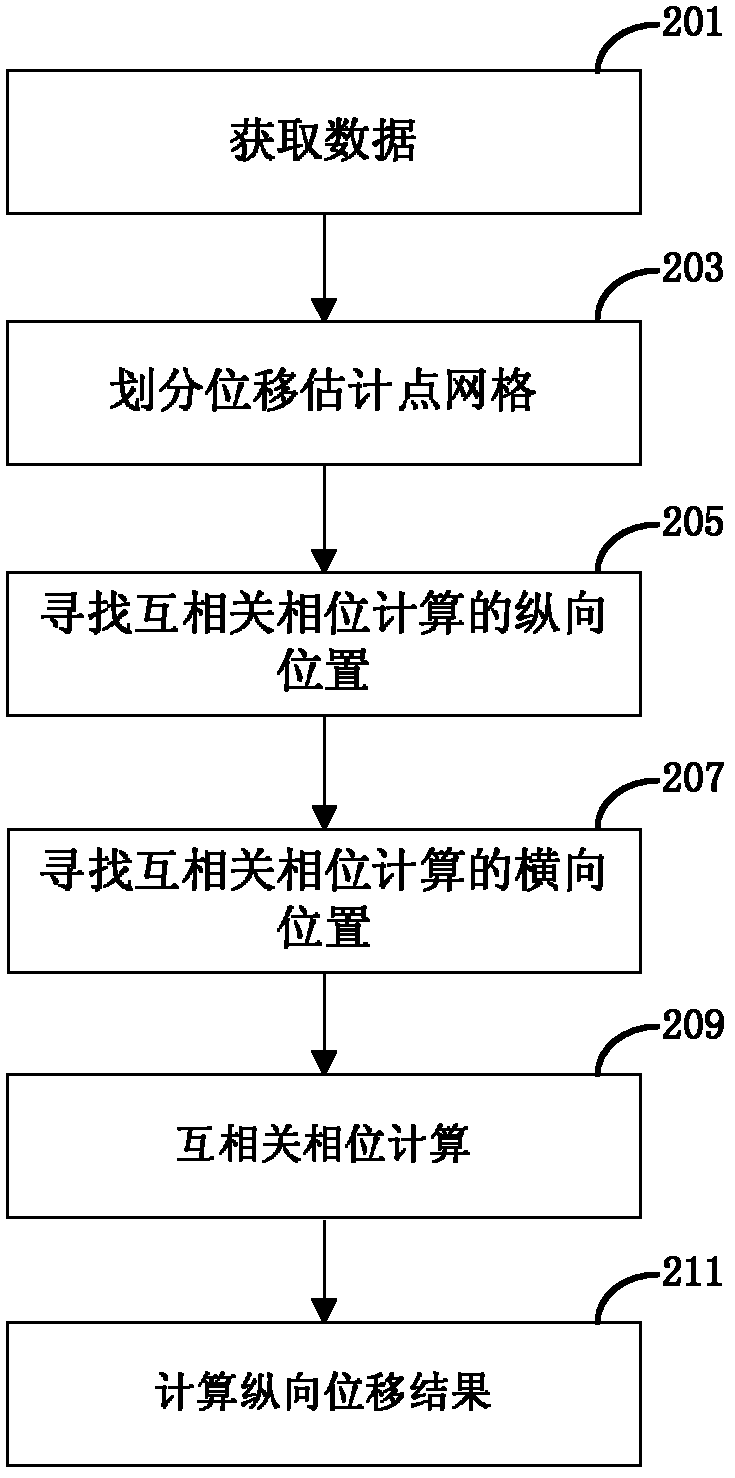
The invention discloses a displacement detecting method and a device thereof in elasticity imaging; the displacement detecting method comprises the following steps: acquiring a target point; acquiring the mutual-correlation phase counting position of the target point in a second frame image; counting a mutual-correlation phase according to the mutual-correlation phase counting position; counting a longitudinal displacement result according to the mutual-correlation phase; and working out a gradient for the displacement result so as to acquire a strain result. According to an elasticity imaging method and a device disclosed by the embodiment of the invention, an two paths of I / Q echo baseband signals which are down-sampled in two frames before and after compression are acquired; displacement information between two frames is detected quickly by guide-type phase estimation; an axial gradient is counted so as to obtain strain information; therefore, a strain image in relatively high quality can be acquired; in addition, counting quantity is reduced greatly; and clinical real-time demands are satisfied.

Description

technical field [0001] The invention relates to a displacement detection method and device in elastic imaging. Background technique [0002] Ultrasound elastography, as an important auxiliary method for the detection of B-mode sonograms in cancer detection, especially in the discrimination of benign and malignant breast cancer, has been rapidly applied in clinic. Ultrasound elastography mainly obtains the ultrasonic echo information of the target tissue through ultrasonic imaging, and then detects the tissue elasticity information through a specific algorithm, and displays it visually in the form of an image to assist doctors in diagnosis or treatment. [0003] The traditional ultrasonic elastography method requires the probe to slightly compress the tissue or use the human body's own breathing, vascular pulsation and other processes to obtain two successive frames of ultrasonic echo signals, and then obtain the displacement between the two frames of signals through a specif...

Claims


Application Information

Patent Timeline
Patent Type & Authority Applications(China)
IPC IPC(8): A61B8/08
CPCG01S7/52017G06T7/60A61B8/485G01N29/0654G01N2291/02827G01S15/8906G01N2291/02475G01S7/52042G06T7/0012
Inventor 李双双樊睿
Owner SHENZHEN MINDRAY BIO MEDICAL ELECTRONICS CO LTD
Learn More