Multi-mode low-coherence scattering spectrometer

A spectrometer and low-coherence technology, which is applied in the field of spectrometer and optical imaging, can solve the problems of insufficient reliability of detection results, lack of quantitative parameters, tissue microstructure, etc., to avoid changes in cell characteristics, convenient control, and simple optical path structure Effect

Inactive Publication Date: 2013-11-27
CHONGQING UNIV
View PDF3 Cites 0 Cited by
  • Summary
  • Abstract
  • Description
  • Claims
  • Application Information

AI Technical Summary

Problems solved by technology

After a series of treatments, the structure or characteristics of the tissue may change, resulting in insufficient reliability of the test results
At the same time, there is also a lack of quantitative parameters in the prior art to describe the changes in tissue microstructure

Method used


Image

  • Multi-mode low-coherence scattering spectrometer

Examples


Embodiment Construction

[0018] The present invention will be described in further detail below in conjunction with the accompanying drawings and specific embodiments.

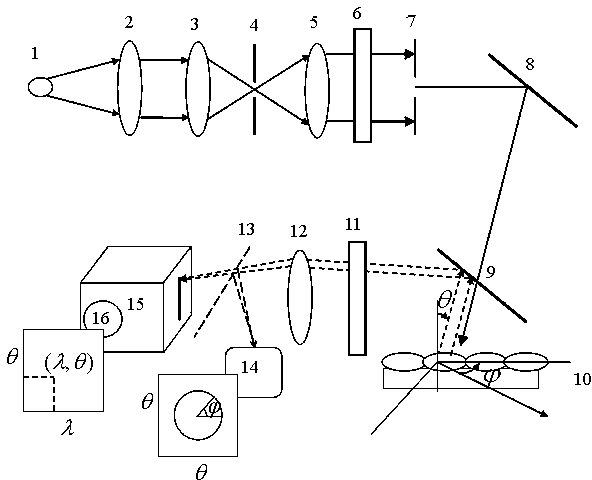
[0019] Such as figure 1 As shown, a multi-mode low-coherence scattering spectrometer is provided with a white light source 1, the white light source 1 adopts a xenon lamp light source, and emits white light with high intensity and low coherence. The white light emitted by the white light source 1 is collimated through the collimation optical path, which is composed of the condenser lens 2, the first lens 3, the first diaphragm 4, the second lens 5 and the second diaphragm 7 in sequence, after collimation The divergence angle of the incident light is 0.3°~0.5°, 0.4° is the best, and the diameter is as low as 2mm.

[0020] A polarizing plate 6 is arranged between the second lens 5 and the second aperture 7, and the linearly polarized light after collimation and polarization is incident on the mirror 8, and the reflected light of the mi...


PUM


Abstract

The invention discloses a multi-mode low-coherence scattering spectrometer, which is characterized by being provided with a white light source, a collimating light path and a polarizer. White light emitted from the white light source irradiates on a reflector after being collimated and polarized, reflected light from the reflector irradiates into an observed sample after transmitting a spectroscope, scattered light of the observed sample is reflected to a polarization analyzer through the spectroscope, light ray permeating the polarization analyzer is absorbed by a duct scope, a spectrometer is mounted along the focal length of the duct scope, a first CCD (charge coupled device) camera is connected to the spectrometer, a movable reflector is arranged between the duct scope and the spectrometer and used for reflecting light ray projected from the duct scope into a second CCD camera. The multi-mode low-coherence scattering spectrometer has the advantages that tissues can be detected noninvasively, variation of cell features caused by dyeing is avoided, structural information of shallow tissue and deep tissue can be acquired simultaneously by the polarization gating technology, spectrum, scattering angle, azimuth angle and polarization character of the tissues can be acquired simultaneously by means of wideband incidence of white light, and structural composition of the tissues can be described quantitatively.

Description

technical field [0001] The invention relates to a spectrometer, in particular to a multi-mode low-coherence scattering spectrometer for tissue cell measurement, which belongs to the optical imaging technology. Background technique [0002] Because traditional histopathological detection often needs to fix the tissue, stain it, and then observe the morphology and structure of the tissue cells through a microscope. A series of treatments may cause changes in the structure or characteristics of the tissue, resulting in insufficient reliability of the test results. At the same time, there is also a lack of quantitative parameters in the prior art to describe changes in tissue microstructures. Therefore, it is of great significance to non-invasively obtain the subdiffuse-scale optical scattering and absorption properties of biological tissue depth tomography to study the structure and composition of tissues. Contents of the invention [0003] The purpose of the present invent...

Claims


Application Information

Patent Timeline
Patent Type & Authority Patents(China)
IPC IPC(8): A61B5/00
Inventor 王品李勇明蒋阳黄敏施忠继邹雪
Owner CHONGQING UNIV
Why Eureka
Try Eureka