Lentivirus vector expression system of luciferase and application thereof
A technology of luciferase and expression system, which is applied in the field of lentiviral vector expression system of luciferase, which can solve the problems of cumbersome process, inability to monitor metastasis, and omission of micrometastases
- Summary
- Abstract
- Description
- Claims
- Application Information
AI Technical Summary
Problems solved by technology
Method used
Image
Examples
Embodiment 1
[0081] Example 1: Construction of a lentiviral vector expression system for luciferase
[0082] Compared with the general plasmid transfection method, the lentiviral expression system has the advantages of high transfection efficiency, stable integration, and continuous expression. Therefore, the inventors constructed a luciferase lentiviral vector expression system. The present inventors replaced the U6 promoter in the FG12 vector in our laboratory with the CMV promoter suitable for eukaryotic expression, and then inserted the improved luciferase reporter gene (Luc2) behind the CMV promoter, while maintaining The original GFP reporter gene module remains unchanged, that is, the FG12 lentiviral vector co-expressing GFP and luciferase is obtained. By co-transfecting 293T cells with the corresponding packaging plasmid, collecting the cell culture supernatant and purifying and concentrating, high-titer virus particles can be obtained for in vivo and in vitro labeling experiments....
Embodiment 2
[0083] Example 2: Establishment of efficient luciferase-labeled tumor tissue method
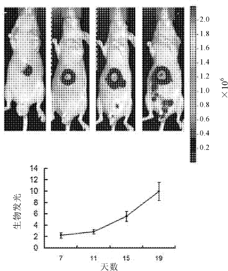
[0084] After the construction of the lentiviral expression system was completed, the inventors marked the liver cancer tissue. For a long time, in vivo labeling of tumor tissue has been a recognized problem, and the relevant literature available for reference is very limited, and some existing reports, such as electroporation, have not achieved satisfactory results. After many attempts and optimizations, the inventors established a more efficient labeling method (see "Materials and Methods"). Experimental data show that this method can effectively infect most of the cells in the tumor tissue block, and the positive rate can reach more than 90%. Moreover, successfully infected tumors can continue to grow in nude mice (including subcutaneous and liver in situ), and become a good model for studying tumor growth, progression, and migration in vivo ( figure 2 ).
[0085] figure 2 Growth of l...
Embodiment 3
[0086] Example 3: Establishment of a nude mouse model for orthotopic transplantation of human liver cancer
[0087] The inventors planted fresh samples of liver cancer on the livers of nude mice in time to establish a nude mouse model of orthotopic transplantation of human liver cancer. Compared with liver cancer models generated from in vitro cell lines, liver cancer models based on patient tumor tissue are closer to the in vivo physiological state of the tumor. In the liver cancer model constructed by the present inventors, the samples used come from liver cancer tissues with a high degree of malignancy, and one of the important features is that the patients have liver portal vein metastasis of liver cancer cells. Therefore, this liver cancer model includes the liver cancer tissue from the primary tumor and the corresponding portal vein tumor thrombus, which can metastasize into blood vessels, and shows a strong ability to metastasize, providing an ideal model for the study ...
PUM
 Abstract
Description
Claims
Application Information
 - R&D
- Intellectual Property
- Life Sciences
- Materials
- Tech Scout
- Unparalleled Data Quality
- Higher Quality Content
- 60% Fewer Hallucinations
Browse by: Latest US Patents, China's latest patents, Technical Efficacy Thesaurus, Application Domain, Technology Topic, Popular Technical Reports.
© 2025 PatSnap. All rights reserved.Legal|Privacy policy|Modern Slavery Act Transparency Statement|Sitemap|About US| Contact US: help@patsnap.com


