
Characterization method of atomic force microscope for micro-pore structure of reservoir rock core

An atomic force microscope and micro-pore technology, applied in the field of oil and gas fields, can solve the problems of inaccuracy, accurate determination of the micro-pore structure error of the reservoir rock, damage to the core structure, etc.

Inactive Publication Date: 2011-09-14
NORTHEAST GASOLINEEUM UNIV
View PDF2 Cites 27 Cited by
  • Summary
  • Abstract
  • Description
  • Claims
  • Application Information

AI Technical Summary

Problems solved by technology

However, the above-mentioned methods have their own advantages and disadvantages in characterizing the microscopic pore structure of reservoir rocks.
Although the on-site evaluation method of well logging data has vertical continuity, due to the influence of various factors such as instruments, environment, fluids, etc., at the same time, there are a lot of well logging data, and there are relatively large human factors in the interpretation, and the macroscopic characteristics of the reservoir can be described. , but its data accuracy and interpretation accuracy cannot be guaranteed for microscopic pore structure research
The capillary pressure curve method in the laboratory method is based on the theory of parallel capillary bundles. It is not suitable for core analysis of fractured and vug reservoirs, and its intuitiveness is also poor.
Only a limited two-dimensional section can be observed in the thin section of the cast body, and all parameters related to the pore structure come from the analysis and calculation of the two-dimensional image, which has certain contingencies and inaccuracies. This will cause secondary mechanical damage, which will damage the inherent structure of the core.
Although scanning electron microscopy has the characteristics of high magnification and large depth of field, it can obtain very clear high-resolution stereoscopic images, and has been widely used in the observation and analysis of microscopic morphology characteristics of rock samples, the division of pore types, and the occurrence of clay minerals. Observation and analysis of phenomena related to filling type, secondary quartz and feldspar, and feldspar dissolution, but the comprehensive effects of secondary electrons, backscattered electrons, transmitted electrons, and differences in the interaction between different material structures and electrons make the obtained images have many In addition, the high-energy electron beam also has a certain destructive effect on the microscopic pore structure of the rock, and although the obtained image is a three-dimensional image, it is difficult to quantify the spatial size. The structure brings more errors
Due to the low resolution of X-CT method and nuclear magnetic resonance method, it is difficult to characterize the micro- and nano-pore structure.

Method used


Image

  • Characterization method of atomic force microscope for micro-pore structure of reservoir rock core
  • Characterization method of atomic force microscope for micro-pore structure of reservoir rock core
  • Characterization method of atomic force microscope for micro-pore structure of reservoir rock core

Examples


Embodiment Construction

[0014] The present invention will be further described below in conjunction with specific examples.

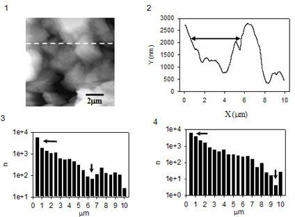
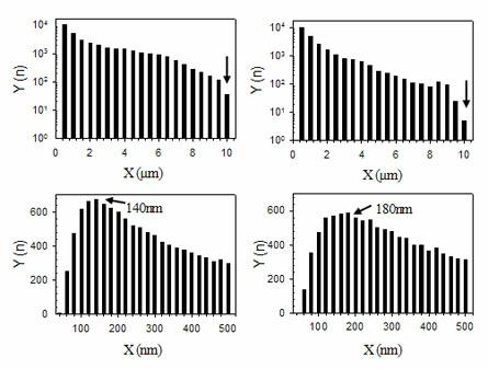
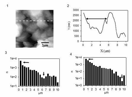
[0015] Firstly, cores were taken industrially at 1138.0 meters from Well Xing2-1-Jian29 in the main oil producing area of ​​Daqing Oilfield, and the rock samples were processed into a cylindrical shape with a diameter of 1 inch on the core bank bench drill, and then core processing equipment was used along the diameter Saw it into thin slices with a thickness of no more than 3 mm, then simply clean the attached dust with an ordinary soft brush and leave it for observation with an atomic force microscope. The actual observed rock samples are core slices with a diameter of 1 inch and a thickness of 3 mm. Note that the surface of the core slices cannot undergo any chemical treatment to ensure the original state of the rock samples. The second is to transform the atomic force microscope to make it suitable for the characterization of the microscopic pore structure of the reservoir...


PUM


Abstract

The invention relates to a characterization method of an atomic force microscope for a micro-pore structure of a reservoir rock core. The method comprises the following steps of: (1) preparing for a characterization sample to ensure that a reservoir rock sample adapts to the characterization of an atomic force microscope; (2) reconstructing a characterization apparatus to ensure that the atomic force microscope adapt to a characterization reservoir rock sample; and (3) analyzing the data of a characterization image, and analyzing the characterization image to obtain useful information to guide production practice. By using the method, the characterization method of the micro-pore structure of the rock can be more complete and accurate so that the characteristics of a nano-grade structure and a micron-grade structure of a reservoir stratum can be indicated three-dimensionally and clearly, and the method has important scientific and economic significances in evaluating reserves of an oil-gas field or the characteristics of the rock micro-pore structure of the oil-gas field to increase the yield of the oil-gas field.

Description

Technical field: [0001] The invention relates to the field of oil and gas fields, and relates to an atomic force microscope characterization method for the microscopic pore structure of reservoir rock cores. Background technique: [0002] At present, the conventional methods for describing the pore structure characteristics of oil and gas reservoir cores mainly include: on-site evaluation method of well logging data and indoor experiment method. The indoor experiment method is currently the most important and the most widely used method to describe and evaluate the characteristics of rock pore structure, mainly including: capillary pressure curve method (semi-permeable diaphragm method, mercury injection method and centrifuge method, etc.), cast thin section scanning electron microscopy, X-CT scanning, and nuclear magnetic resonance. However, the above-mentioned methods have their own advantages and disadvantages in characterizing the microscopic pore structure of reservoir...

Claims


Application Information

Patent Timeline
IPC IPC(8): G01N15/08G01Q60/24
Inventor 白永强张雁张北虹赵荣
Owner NORTHEAST GASOLINEEUM UNIV
Learn More