Magnetic resonance imaging apparatus

A magnetic resonance and imaging technology, applied in magnetic resonance measurement, measurement using nuclear magnetic resonance image system, measurement equipment, etc., can solve the problems that coronary arteries take time and reliability depends on doctors, etc.

Active Publication Date: 2011-07-20
TOSHIBA MEDICAL SYST CORP
View PDF7 Cites 2 Cited by
  • Summary
  • Abstract
  • Description
  • Claims
  • Application Information

AI Technical Summary

Problems solved by technology

Therefore, there is a problem that it takes time to set the resting period of the coronary artery, and the reliability of the set resting period depends on the skill level of the physician.

Method used


Image

  • Magnetic resonance imaging apparatus
  • Magnetic resonance imaging apparatus
  • Magnetic resonance imaging apparatus

Examples


Embodiment Construction

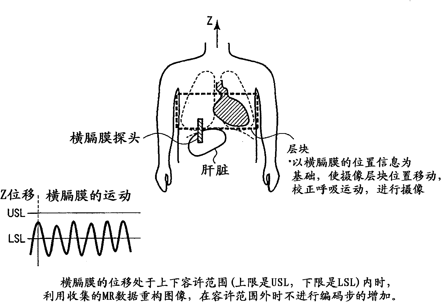
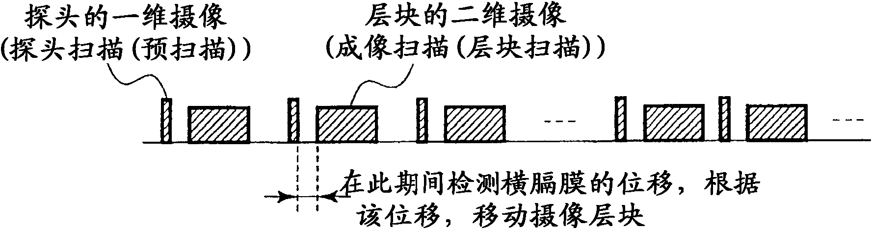
[0030] In general, according to one embodiment, the magnetic resonance imaging device repeatedly performs imaging scans of the entire heart of the subject, and performs probe scans immediately before each of the above-mentioned imaging scans, and detects the lateral movement caused by the respiratory movement of the subject. The displacement of the diaphragm is detected, and the imaging range of each imaging scan is shifted based on the detected displacement of the diaphragm. In order to generate a series of cross-sectional images by synchronously repeating electrocardiographic imaging of the area including the heart as a preparatory scan for the probe scan and imaging scan, the control unit controls the transmitter and receiver unit of the RF coil and the gradient magnetic field power supply. Using a series of cross-sectional images as objects, the first static period in which the positional fluctuations of the coronary arteries converge within a certain range during the cardi...


PUM


Abstract

The invention provides a magnetic resonance imaging apparatus capable of displacing an imaging range for each of the imaging scans based on the detected displacement of the diaphragm. A control unit controls an RF coil reception / transmission unit and a gradient field power supply to generate a series of slice images by repeatedly imaging a region including the heart by ECG gating for preliminary scans for the probe scans and the imaging scans. This apparatus specifies, for the series of slice images, the first rest period in which a variation in position of the coronary artery falls within a predetermined range in a cardiac cycle, based. on a change in the image of the overall heart. The apparatus specifies the second rest period of the coronary artery in the cardiac cycle, for a plurality of slice images, of the series of slice images, which correspond to the specified first rest period or a rest period enlarged from the first rest period, by tracking the movement of the coronary artery only within a local range including the coronary artery. The apparatus then reconstructs an image based on MR data acquired in the second rest period.

Description

[0001] Cross References to Related Applications [0002] This application is based on and claims priority from prior Japanese Patent Application No. 2010-010412 filed on January 20, 2010, the entire contents of which are incorporated herein by reference. technical field [0003] The embodiments described here relate generally to a magnetic resonance imaging system. technical background [0004] In the case of imaging (Whole Heart MRCA (WHMRCA): Whole Heart Magnetic Resonance Coronary Angiography) that visualizes the movement of the coronary arteries, particularly the coronary arteries of the entire heart, using a magnetic resonance imaging device (MRI), using a three-dimensional The ssfp (steady state free precession: steady state free precession) sequence is a method of taking pictures under natural respiration. At this time, the motion of the heart caused by breathing motion is corrected by the RMC (Real-time motion correction) method, and an image is taken. On the other...

Claims


Application Information

Patent Timeline
IPC IPC(8): A61B5/055
CPCG01R33/56509G01R33/5676G06T2207/10088G01R33/5635G01R33/56325G06T7/20G01R33/48G06T2207/20016G06T7/0038G06T2207/30048A61B5/055
Inventor 久原重英二宫绫子
Owner TOSHIBA MEDICAL SYST CORP
Why Eureka
Try Eureka