Application of Cucurmosin in preparation of medicaments for treating cervical cancer
A technology for cervical cancer and pumpkin is applied in the application field of pumpkin protein in the preparation of antitumor drugs, and can solve problems such as damage to normal tissues and cells.
- Summary
- Abstract
- Description
- Claims
- Application Information
AI Technical Summary
Problems solved by technology
Method used
Image
Examples
Embodiment 1
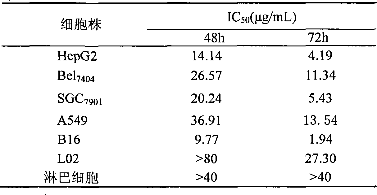
[0011] Example 1: Inhibitory effect of pumpkin protein on the proliferation of tumor cells cultured in vitro and its influence on human normal cells
[0012] Tumor cells in the logarithmic growth phase (gastric cancer cell SGC 7901 , HepG 2 、Bel 7404 , non-small cell lung cancer cell A 549 , breast cancer cells MCF-7 and melanoma cells B16) and normal human liver cells L02 were inoculated on a 96-well culture plate, and the inoculum volume per well was 3000-4000 cells / 100 μL and 6000 cells / 100 μL respectively; healthy blood donations were collected aseptically 15 mL of venous blood from patients, anticoagulated with heparin, separated mononuclear cells by Ficoll density gradient centrifugation, prepared single cell suspension with RPMI1640 containing 10% fetal bovine serum, and adjusted the cell concentration to 2×10 5 cells / mL, inoculated in 96-well culture plate, 100 μL per well. After 24 hours of cell culture on the above-mentioned culture plates, 100 μL of pumpkin prot...
Embodiment 2
[0015] Example 2: Pumpkin protein to SGC 7901 , HepG 2 、Bel 7404 、A 549 , MCF-7 and B16 cells inducing apoptosis
[0016] (1) Effect of pumpkin protein on the morphology of the above tumor cells
[0017] ①Fluorescence microscope observation: the above tumor cells were inoculated on multiple coverslips, treated with different concentrations of pumpkin protein for 24, 48 and 72 hours after 12 hours, and stained with acridine orange (AO) / ethidium bromide (EB) mixed fluorescent dyes Afterwards, place it upside down on a glass slide, and observe the morphological changes of the cells under a fluorescence microscope. Results: The above cells can be observed in the nucleus or cytoplasm of apoptotic cells in the early and middle stages, with densely stained granular yellow-green fluorescence, nuclear chromatin condensation, nuclear rupture, and the release of apoptotic bodies. Typical apoptotic cell morphology There are time- and dose-dependent changes in apoptosis.
[0018] ② E...
Embodiment 3
[0022] Example 3: Induction and differentiation of pumpkin protein on tumor cells (using melanoma B16 cells as a model)
[0023] (1) Observation of cell morphology: After the B16 cells in the control group and the experimental group were cultured for 72 hours, the cell morphology was observed and photographed with a phase contrast microscope and a transmission electron microscope, respectively. The result is as follows:
[0024] ①Observation by optical microscope: B16 cells in the control group grew irregularly in multiple layers, with overlapping round cells; after the treatment of pumpkin protein, the cells grew in a certain polarity, arranged in parallel without overlapping; after 3 days, the concentration of pumpkin protein was 2.5 For those above μg / mL, the polarity of the cells is obvious, most of the cells become larger, have a dendritic structure, the cells are connected into a network, grow slowly, and the number of cells decreases.
[0025] ② Electron microscope obser...
PUM
 Abstract
Description
Claims
Application Information
 - Generate Ideas
- Intellectual Property
- Life Sciences
- Materials
- Tech Scout
- Unparalleled Data Quality
- Higher Quality Content
- 60% Fewer Hallucinations
Browse by: Latest US Patents, China's latest patents, Technical Efficacy Thesaurus, Application Domain, Technology Topic, Popular Technical Reports.
© 2025 PatSnap. All rights reserved.Legal|Privacy policy|Modern Slavery Act Transparency Statement|Sitemap|About US| Contact US: help@patsnap.com


