Method for staining plant tissue section by using thionine
A plant tissue and slice dyeing technology, applied in the field of plant slice dyeing, can solve the problems of single color, influence of observation effect, inability to distinguish different tissues, etc., and achieve intuitive effect
- Summary
- Abstract
- Description
- Claims
- Application Information
AI Technical Summary
Problems solved by technology
Method used
Image
Examples
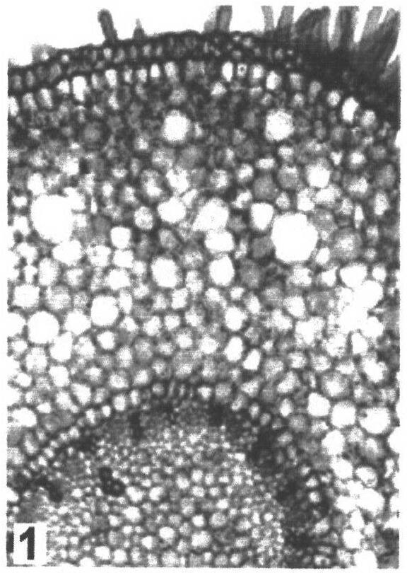
Embodiment 1
[0015] (1) 30 milliliters of thionine solutions with a mass fraction of 0.2% are prepared, and added to a petri dish for use;
[0016] (2) Get the root of Chlorophytum chinensis with root hairs, slice it by hand and store it in a petri dish filled with clear water;
[0017] (3) Dip the plant tissue section soaked in clear water in (2) with a brush, and transfer to the petri dish containing the thionine solution in (1) for staining for 3 minutes;
[0018] (4) Dip the stained plant tissue section in (3) with a brush, transfer it to a glass slide, and drop a drop of 0.5% NaHCO on the section 3 Solution, cover the cover glass, gently press the cover glass with tweezers, then absorb excess water around the cover glass with absorbent paper, observe and take pictures under the microscope.
[0019] As shown in Figure 1, the root hair cells and epidermal cells of the root of Chlorophytum are purple, the parenchyma cells of the cortex are mostly light pink, the endothelial cells are li...
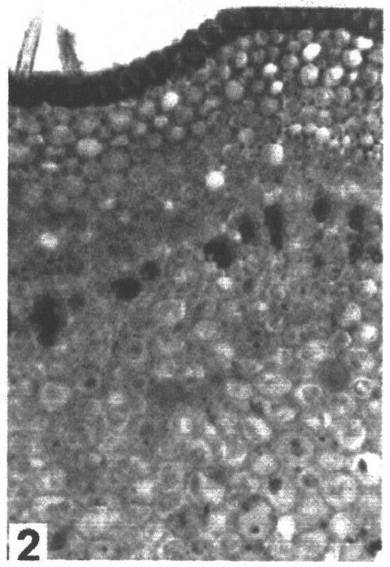
Embodiment 2
[0021] (1) 30 milliliters of thionine solutions with a mass fraction of 0.5% are prepared, and added to a petri dish for use;
[0022] (2) Get the stem of Euonymus microphylla, cut the plant material by free-hand slicing, and store the slices in a petri dish filled with clear water for later use;
[0023] (3) Dip the plant tissue section soaked in clear water in (2) with a brush, and transfer to the petri dish containing the thionine solution in (1) for staining for 5 minutes;
[0024] (4) Dip the stained plant tissue section in (3) with a brush, transfer it to a glass slide, and drop a drop of 1% NaHCO on the section 3 Solution, cover the cover glass, gently press the cover glass with tweezers, then absorb excess water around the cover glass with absorbent paper, observe and take pictures under the microscope.
[0025] As shown in Figure 2, the cell wall of the stem epidermal cells of Euonymus microphylla is blue, the epidermal hairs are colorless, the vessels are blue, and ...
PUM
 Abstract
Description
Claims
Application Information
 - R&D Engineer
- R&D Manager
- IP Professional
- Industry Leading Data Capabilities
- Powerful AI technology
- Patent DNA Extraction
Browse by: Latest US Patents, China's latest patents, Technical Efficacy Thesaurus, Application Domain, Technology Topic, Popular Technical Reports.
© 2024 PatSnap. All rights reserved.Legal|Privacy policy|Modern Slavery Act Transparency Statement|Sitemap|About US| Contact US: help@patsnap.com


