
Hard ultrasonic gallbladder endoscope system and using method thereof

A hard and gallbladder technology, applied in the direction of endoscopy, application, laparoscopy, etc., can solve the problems of weakened optical signal, insufficient diagnostic accuracy, fiber optic restriction and long transmission distance, etc., to achieve enhanced grasp and clear observation and the effect of processing

Active Publication Date: 2010-05-12
GUANGZHOU BAODAN MEDICAL INSTR TECH
View PDF0 Cites 7 Cited by
  • Summary
  • Abstract
  • Description
  • Claims
  • Application Information

AI Technical Summary

Problems solved by technology

[0002] At present, the existing biliary endoscopy used for gallbladder disease surgery can determine the lesion in the gallbladder cavity, but it cannot understand the changes of the layers of the gallbladder wall and surrounding organs, and there is a hidden danger of insufficient diagnostic accuracy
At present, in clinical medicine, the understanding of gallbladder lesions mainly relies on in vitro B-ultrasound and CT technology, which cannot provide accurate information on the lesions of each layer of the gallbladder wall and the organs around the gallbladder.
In addition, the current ultrasonic endoscopic technology has been widely used in gastric, duodenal and other surgical fields, and its equipment is a variety of soft mirrors and supporting miniature ultrasonic probes, etc., but it has not been used in the diagnosis and treatment of gallbladder
The early gallbladder-preserving stone removal and polyp surgery used soft fiber choledochoscope. When the doctor used the soft fiber choledochoscope to perform the operation, because the end that enters the human body is soft, it cannot transmit the force well, and it is different from the hard gallbladder. Compared with endoscopes, the maneuverability is poor; because the optical system of the flexible fiber choledochoscope uses fiber optic image transmission, and the fiber optic itself is restricted and the transmission distance is long, the optical signal will gradually weaken during the image transmission process, and its imaging The display effect of the system is not as good as that of the rigid endoscope that uses cylindrical lens imaging, which increases the difficulty of the doctor's operation, and cannot control the flexible fiber choledochoscope well for surgery, resulting in stones and polyps remaining in the gallbladder

Method used


Image

  • Hard ultrasonic gallbladder endoscope system and using method thereof
  • Hard ultrasonic gallbladder endoscope system and using method thereof
  • Hard ultrasonic gallbladder endoscope system and using method thereof

Examples


Embodiment Construction

[0025] Below in conjunction with accompanying drawing, the present invention is described in further detail:

[0026] The invention comprises a rigid gallbladder endoscope with a rigid mirror body, a miniature ultrasonic probe, an external camera system and an external light source system connected with the rigid gallbladder endoscope and the miniature ultrasonic probe.

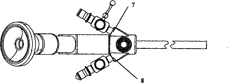
[0027] Such as figure 1 , figure 2 As shown, the rigid gallbladder endoscope of the present invention is composed of instrument channel 1, eyepiece input end 2, endoscope main body 3, cold light source input end 4, endoscope main body front 5, endoscope end 6, water inlet channel 7 and The water outlet channel 8 is composed of the instrument channel 1, the water inlet channel 7, the water outlet channel 8, the eyepiece input end 2, and the cold light source input end 4. , endoscope end 6 . In the present invention, the cold light source input end 4 is designed to form an included angle of 90 degrees with ...


PUM


Abstract

The invention belongs to the field of medical apparatus, and particularly relates to a hard ultrasonic gallbladder endoscope system. The hard ultrasonic gallbladder endoscope system comprises a hard gallbladder endoscope with a hard endoscope body, a miniature ultrasonic probe, and an external camera system and an external light source system which are connected with the hard gallbladder endoscope and the miniature ultrasonic probe. The hard gallbladder endoscope of the hard ultrasonic gallbladder endoscope system enters a gallbladder cavity through a micro incision at the bottom of a gallbladder; the miniature ultrasonic probe enters the gallbladder directly through an instrument channel of the hard gallbladder endoscope under the monitoring of the camera system of the hard gallbladder endoscope by taking the hard gallbladder endoscope as a platform, and simultaneously the miniature ultrasonic probe directly observes the condition in the gallbladder cavity clearly; and under the condition that the gallbladder is filled with liquid, the hard gallbladder endoscope makes diagnoses and gives treatment to various gallbladder diseases, the miniature ultrasonic probe performs real-time ultrasonic scanning and performs circular scanning, linear scanning or three-dimensional reconstruction in 360 degrees on the cavity so as to acquire change images of each layer of the gallbladder wall and organs surrounding the gallbladder, so the hard ultrasonic gallbladder endoscope system can determine the position, the size, the appearance and the range of a focus of a disease, improves the medical quality and the medical safety, and overcomes the defects of a diagnosis and treatment method at present.

Description

technical field [0001] The invention belongs to the field of medical instruments, in particular to a rigid ultrasonic gallbladder endoscope system for endoscopic gallbladder disease surgery. Background technique [0002] At present, the existing biliary endoscopy used for gallbladder disease surgery can determine the lesion in the gallbladder cavity, but it cannot understand the changes of the layers of the gallbladder wall and the surrounding organs, and there is a hidden danger of insufficient diagnostic accuracy. At present, in clinical medicine, the understanding of gallbladder lesions mainly relies on in vitro B-ultrasound and CT techniques, which cannot provide accurate information on the lesions of each layer of the gallbladder wall and the organs around the gallbladder. In addition, the current endoscopic ultrasonography technology has been widely used in gastric, duodenal and other surgical fields, and its equipment is various soft mirrors and supporting micro-ultra...

Claims


Application Information

Patent Timeline
Patent Type & Authority Applications(China)
IPC IPC(8): A61B17/22A61B1/313A61B17/94A61B1/04A61B1/06A61B8/12
Inventor 乔铁
Owner GUANGZHOU BAODAN MEDICAL INSTR TECH
Learn More