
Method for automatically focusing microscope system in urinary sediment examination equipment

A microscope system and testing equipment technology, applied in biochemical equipment and methods, microscopes, enzymology/microbiology devices, etc., can solve problems such as flow microscope systems that cannot be directly applied to urine sediment testing equipment, and meet the requirements. , Focusing judgment is accurate, focusing is accurate

Active Publication Date: 2012-01-25
DIRUI MEDICAL TECH CO LTD
View PDF0 Cites 0 Cited by
  • Summary
  • Abstract
  • Description
  • Claims
  • Application Information

AI Technical Summary

Problems solved by technology

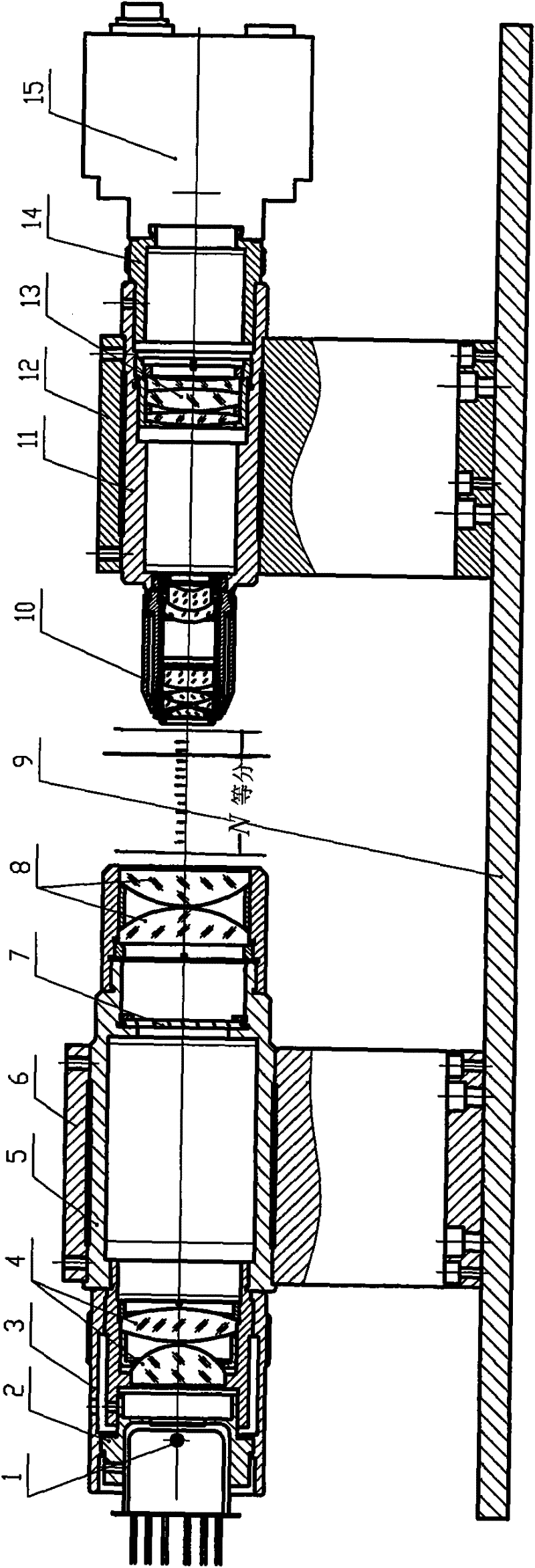
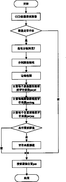
[0005] The invention provides an automatic focusing method for a microscope system in urine sediment testing equipment to solve the problem that none of the existing automatic focusing methods can be directly applied to the flow microscope system of urine sediment testing equipment

Method used


Image

  • Method for automatically focusing microscope system in urinary sediment examination equipment
  • Method for automatically focusing microscope system in urinary sediment examination equipment
  • Method for automatically focusing microscope system in urinary sediment examination equipment

Examples


Embodiment Construction

[0024] Glossary:

[0025] Urinary sediment: Refers to the formed components in urine, such as red blood cells, white blood cells and bacteria in urine.

[0026] Urine sediment testing equipment: It is a clinical testing equipment for detecting formed components in urine.

[0027] Focusing fluid: a liquid containing solidified red blood cells, generally at a concentration of about: 0.8 x 10 6 ~1.5×10 6 pcs / ul, used for the automatic focusing process of the microscope system in the urine sediment testing equipment.

[0028] Focusing Particles: Solidified red blood cells in focusing fluid.

[0029] Laminar flow: Laminar flow refers to the orderly flow of fluid microgroups without mixing with each other.

[0030] Flow cell: It is composed of a specially made thin-layer plate, and the detection sample forms a laminar flow under the action of the sheath fluid.

[0031] Include the following steps:

[0032] (1) If figure 1 As shown, the focusing interval is divided into N=500 ...


PUM


Abstract

The invention relates to a method for automatically focusing a microscope system in urinary sediment examination equipment, belonging to method for processing the image of the microscope system. Firstly, the image of focusing liquid passing by a flow cell in a laminar flow way at high speed is shot, and then the image of the focusing liquid is analyzed to find the optimal focusing position and automatically focus the microscope system in the urinary sediment examination equipment. Aiming at solving the practical problem of automatically focusing the microscope system in the urinary sediment examination equipment, the invention provides a novel method for automatically focusing the microscope system in the urinary sediment examination equipment. The novel method can accurately focus the microscope system in urinary sediment examination equipment and has good repeatability. The invention provides the method for calculating the evaluation function of the definition of the image in each position on the basis of calculating the definition of the focusing particles in the focusing liquid, thereby better meeting the requirement of the evaluation function of the definition of the image and making the focusing judgment more accurate.

Description

technical field [0001] The invention belongs to the technical field of image processing of microscope systems, in particular to an automatic focusing method in a urine sediment microscope system. Background technique [0002] In terms of basic principles, automatic focusing methods can be divided into two categories: one is the active method; the other is the passive method. The active method is a ranging method based on the distance measurement between the lens and the object to be photographed. The optical system is adjusted according to the situation of the object to be photographed. The characteristic of the active method is to measure the distance and orientation of the object by receiving the reflection of the actively emitted electromagnetic wave or sound wave. And calculate the best focus position according to the principle of optical imaging, and then use the information processor to control the motor to achieve automatic focus, so this method is called active metho...

Claims


Application Information

Patent Timeline
Patent Type & Authority Patents(China)
IPC IPC(8): G02B21/24C12M1/34G01N21/84
Inventor 宋洁沈继楠范洪岩
Owner DIRUI MEDICAL TECH CO LTD
Learn More