Non-contact type optical sectioning imaging method

An optical tomography, non-contact technology, used in diagnosis, medical science, diagnostic recording/measurement, etc., can solve problems such as the inability to achieve non-contact optical tomography

Inactive Publication Date: 2010-04-14
XIDIAN UNIV
View PDF0 Cites 27 Cited by
  • Summary
  • Abstract
  • Description
  • Claims
  • Application Information

AI Technical Summary

Problems solved by technology

However, this method is only applicable to the case of small-aperture lenses, and is not suitable for slightly larger-aperture lenses...

Method used


Image

  • Non-contact type optical sectioning imaging method
  • Non-contact type optical sectioning imaging method
  • Non-contact type optical sectioning imaging method

Examples


Embodiment Construction

[0037] The reconstruction method of the present invention will be described in detail below in conjunction with the accompanying drawings. It should be noted that the described embodiments are only intended to facilitate the understanding of the present invention, and have no limiting effect on it.

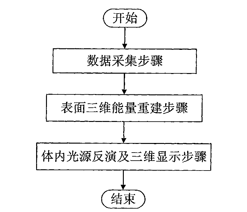
[0038] refer to figure 1 , the non-contact optical tomography of the present invention comprises the following steps:

[0039] Step 1, collecting multi-angle optical images and biological surface shape and anatomical structure data.

[0040] (1.1) Build a multimodal optical molecular imaging system, which includes two subsystems: a non-contact optical tomography system and a microcomputer tomography system. Among them, the non-contact optical tomography system is composed of a high-performance CCD camera and imaging lens, which is used to collect multi-angle two-dimensional optical images; the micro-computed tomography system is composed of an X-ray emission tube and an X-ray det...


PUM


Abstract

The invention discloses a non-contact type optical sectioning imaging method, and solves the problem of the non-contact type optical sectioning imaging which can not be realized in the prior art under high-aperture lenses. The method comprises the following realization steps: firstly, using a multimode optical molecule imaging system to collect multi-angle optical images, organism surface three-dimensional shapes and anatomical structure information, then, reconstructing organism surface three-dimensional energy, including: (1) preprocessing optical images and conversing from gray value to energy value, (2) registering the optical images and organism surface three-dimensional shape information, (3) confirming virtual detecting surface position and pixel size, and mapping energy of CCD cameras to the virtual detecting surface, (4) computing visible factors, (5) reconstructing surface three-dimensional energy distribution, finally, inverting the internal light source, and displaying three-dimensionally and solidly. The method has the advantages of realizing surface three-dimensional energy reconstruction under high-aperture lenses and non-contact type sectioning imaging under high-aperture lenses, and can be used in the field of non-contact type optical molecule imaging.

Description

technical field [0001] The invention belongs to the technical field of optical molecular imaging, and relates to a non-contact optical tomographic imaging technology, in particular to an imaging method for indirect information from a multi-angle two-dimensional optical image measured by a non-contact CCD camera to an internal light source of a living body. Background technique [0002] In recent years, optical molecular imaging technology has been widely used in small animal research due to its high sensitivity, low price and easy operation. In order to obtain an intuitive three-dimensional visualization imaging effect, the newly developed optical tomography imaging technology of optical molecular imaging technology has become a new application research hotspot. In vivo tissue lesion information. In optical tomography, excited fluorescence tomography and bioluminescence tomography are two main imaging modalities. [0003] Optical tomography technology reconstructs the ligh...

Claims


Application Information

Patent Timeline
IPC IPC(8): A61B5/00
Inventor 田捷陈雪利梁继民屈晓超高新波赵恒王琳刘俊廷
Owner XIDIAN UNIV
Why Eureka
Try Eureka