Contact type eyeground imaging system

An imaging system and contact technology, applied in fundus mirrors, eye testing equipment, medical science, etc., can solve the problems of small fundus scope, expensive equipment, unfavorable patient data preservation, analysis and follow-up, etc., to avoid corneal epithelium drying. , Easy to operate, friendly software interface

Inactive Publication Date: 2009-07-08
北京宏仁凝瑞科技发展有限公司
View PDF1 Cites 11 Cited by
  • Summary
  • Abstract
  • Description
  • Claims
  • Application Information

AI Technical Summary

Problems solved by technology

[0012] The disadvantages are: ① Electricity is needed as a light source, ② The range of fundus that can be seen is small
The advantage of this method is that the cost of equipment is relatively low, and it is convenient to carry out in hospitals at all levels. However, it has the following disadvantages: 1. It usually takes several months to master the correct use of binocular indirect ophthalmoscope and the accurate diagnosis of fundus images. For the less common ROP, the learning cycle may be longer; 2. Newborns do not cooperate during the examination, and the twisting of their body, especially the head, increases the difficulty and time of the examination; 3. Due to the regularity of retinal blood vessel development, Most ROP lesions are located in the peripheral retina, and their inspection requires high skills and experience, which is easy to be missed; 4. The cornea is exposed during the inspection process, which may cause corneal epithelial edema; 5. The lack of objective records is not conducive to the collection of patient data Preservation, further condition analysis and follow-up
[0019] The disadvantage is that the camera is installed in the host and connected to the camera through an optical cable. The equipment is too large and inconvenient to move; multiple images are needed to see the peripheral retina clearly; because the control parts are not centralized, the operation requires two people to cooperate to complete; equipment price Expensive, unaffordable for hospitals in remote areas, low penetration rate, patients can only choose a few well-known large domestic hospitals for examination, increasing the burden on patients

Method used


Image

  • Contact type eyeground imaging system
  • Contact type eyeground imaging system
  • Contact type eyeground imaging system

Examples


Embodiment Construction

[0040] The following examples are used to illustrate the present invention, but are not intended to limit the scope of the present invention.

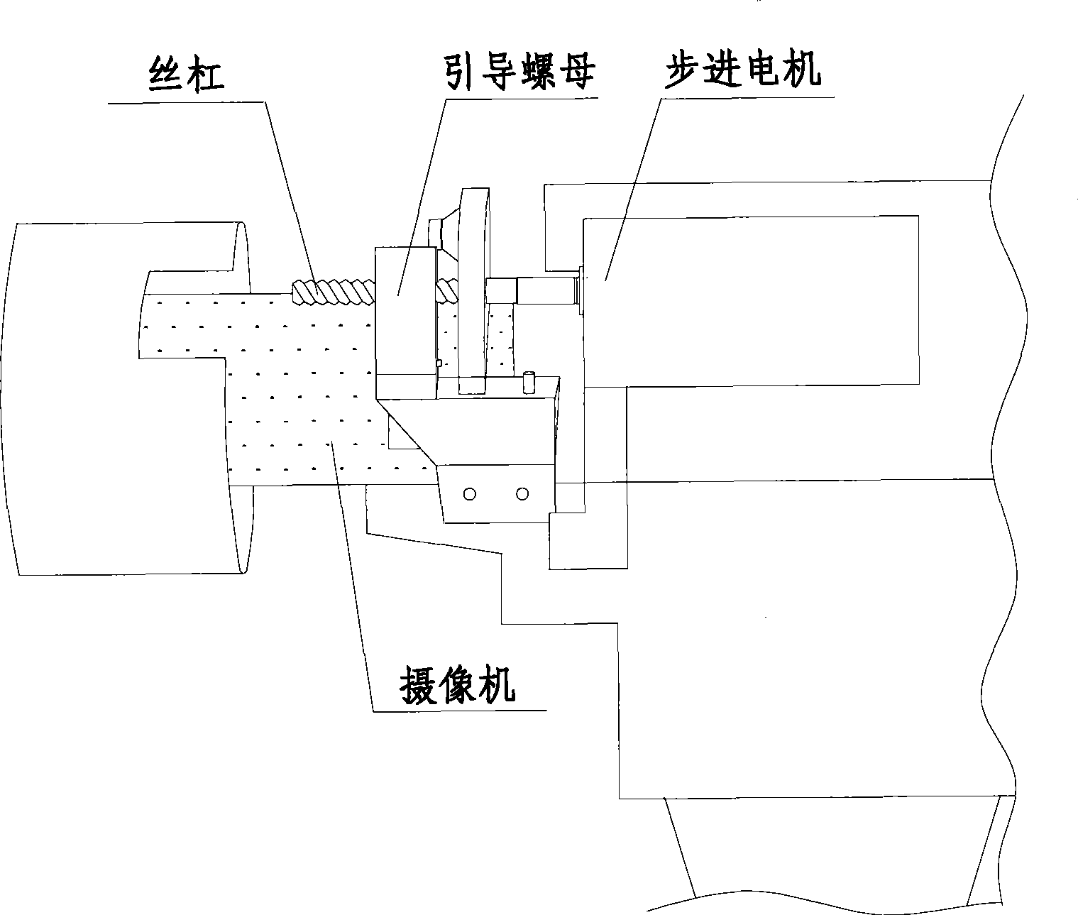
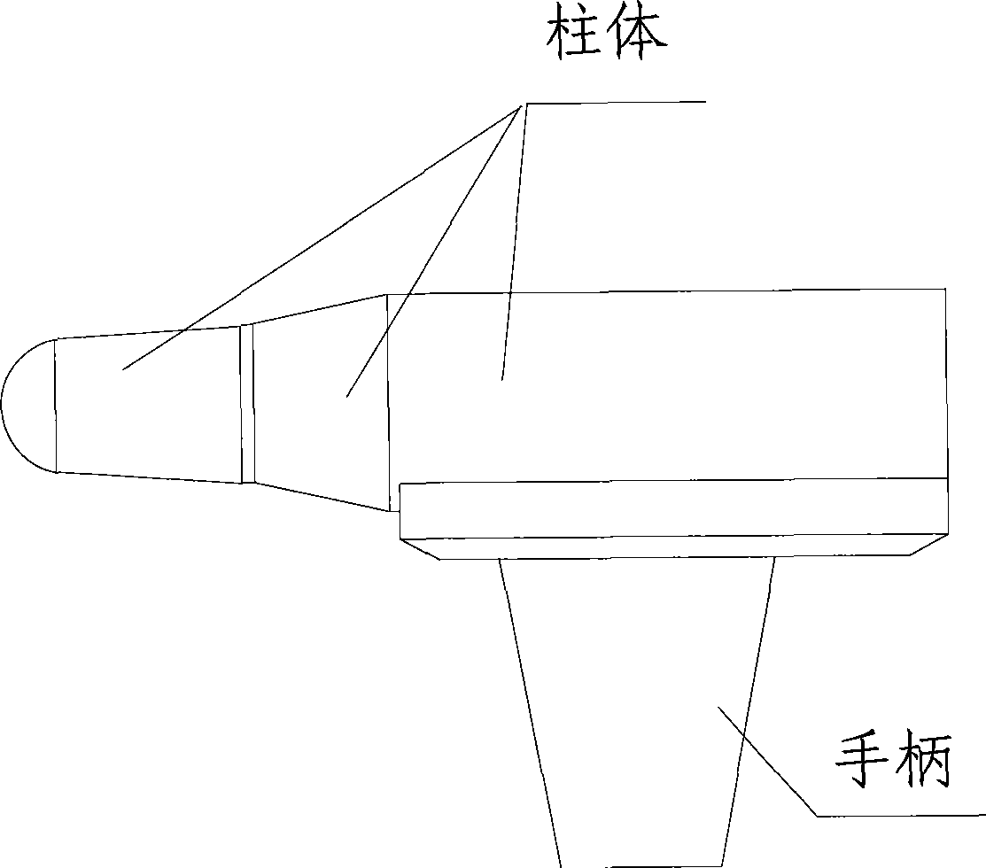
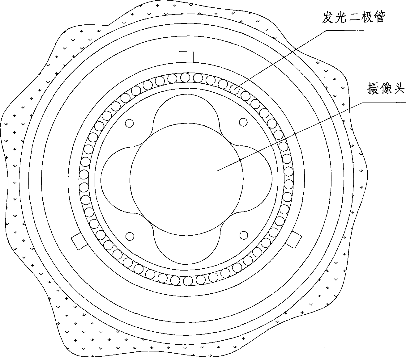
[0041] as attached Figures 1 to 5 As shown, a specific embodiment of a contact fundus imaging system of the present invention is as follows. The system is connected together by a digital camera and a computer through a wired or wireless network, and a coupling agent "transparent gel" is sandwiched between the camera unit and the eyeball. Glue", the digital camera is used to capture fundus images. The digital camera is installed in a cylinder, a stepping motor is also installed in the cylinder, a guide screw and a guide nut are installed at the front end of the stepping motor, and the camera unit and the guide The nuts are connected together, and the stepping motor drives the guide screw to rotate, so that the guide nut moves back and forth to adjust the focal length of the camera head; one end of the cylinder gradually becomes thinne...


PUM


Abstract

The invention relates to an apparatus for medical examination, and provides a contact eyeground imaging system. The system comprises a camera unit and a computer which are connected by a wire or wireless network, and the camera unit is used for shooting eyeground images. The camera unit is arranged in a column one end of which gradually becomes thinner and thinner, the top end is a sphere, and the camera unit is arranged in the sphere. LEDs are arranged around the camera unit in the column. One side of the column is equipped with a focusing button, a dimmer button, and a camera switch button, and clear gel is clamped between the camera unit and an eyeball. Size and shape of the top sphere at one end of the column are fit for human eyeballs. The system has the advantages of wide examination range of the periphery of the eyeground for the examination device and high resolution of pictures, and the operation can be finished independently by one person or by the cooperation of two persons. The examination process is simple, thus discomfort of the persons to be examined is less than that of the persons to be examined by the normal method.

Description

technical field [0001] The invention relates to an instrument for medical examination, in particular to an instrument for examining eyes. Background technique [0002] At present, conventional fundus examination at home and abroad is to use ophthalmoscope to check the back of the eyeball such as vitreous, retina, choroid and optic nerve head. The inspection is generally carried out in a dark room. The principle (take the reflective ophthalmoscope as an example) is mainly to use the ophthalmoscope to irradiate light into the subject's eyes through the pupil, and the light reflected by the fundus of the subject is imaged on the set. The light microscope and those in front of the examiner's eyes are called indirect inspection methods, and those whose images are in the examiner's eyes are called direct inspection methods. Due to the use of the ophthalmoscope, not only can check the various tissues in the eye, such as the optic nerve, retina, choroid, and the transparent tissues...

Claims


Application Information

Patent Timeline
Patent Type & Authority Applications(China)
IPC IPC(8): A61B3/125A61B3/14
Inventor 苏志宏
Owner 北京宏仁凝瑞科技发展有限公司
Why Eureka
Try Eureka