
Method for detecting lymphocyte subgroup with mono-clone antibody SPA hematid rosette method

A monoclonal antibody and lymphocyte technology, applied in the field of medical immunology, can solve problems such as loss, difficulty, and inability to adapt, and achieve the effect of reducing systematic errors and accidental errors, avoiding time-consuming and complicated operations, and reducing cell damage

Inactive Publication Date: 2011-01-26
甘肃省医学科学研究院
View PDF0 Cites 0 Cited by
  • Summary
  • Abstract
  • Description
  • Claims
  • Application Information

AI Technical Summary

Problems solved by technology

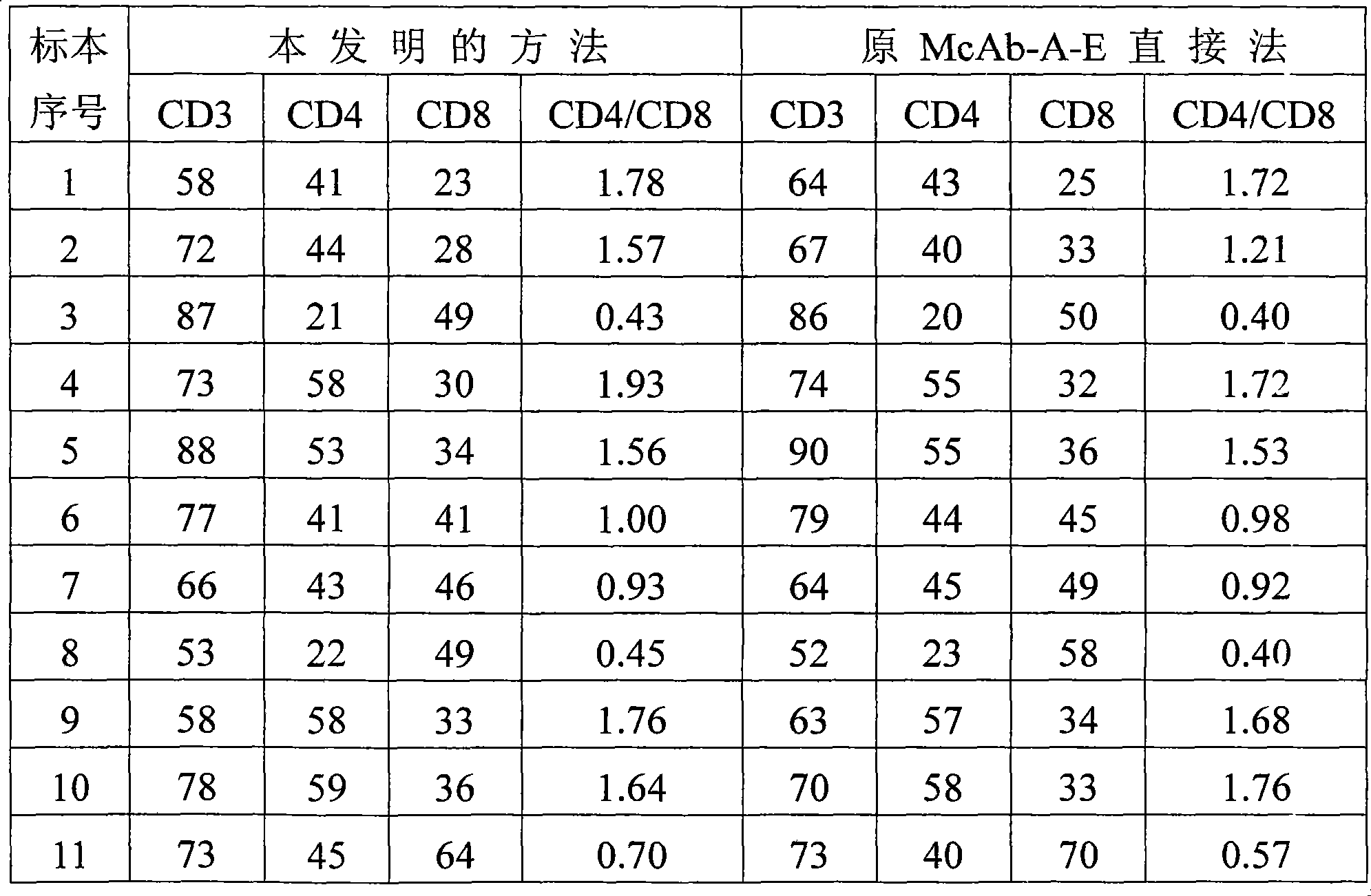
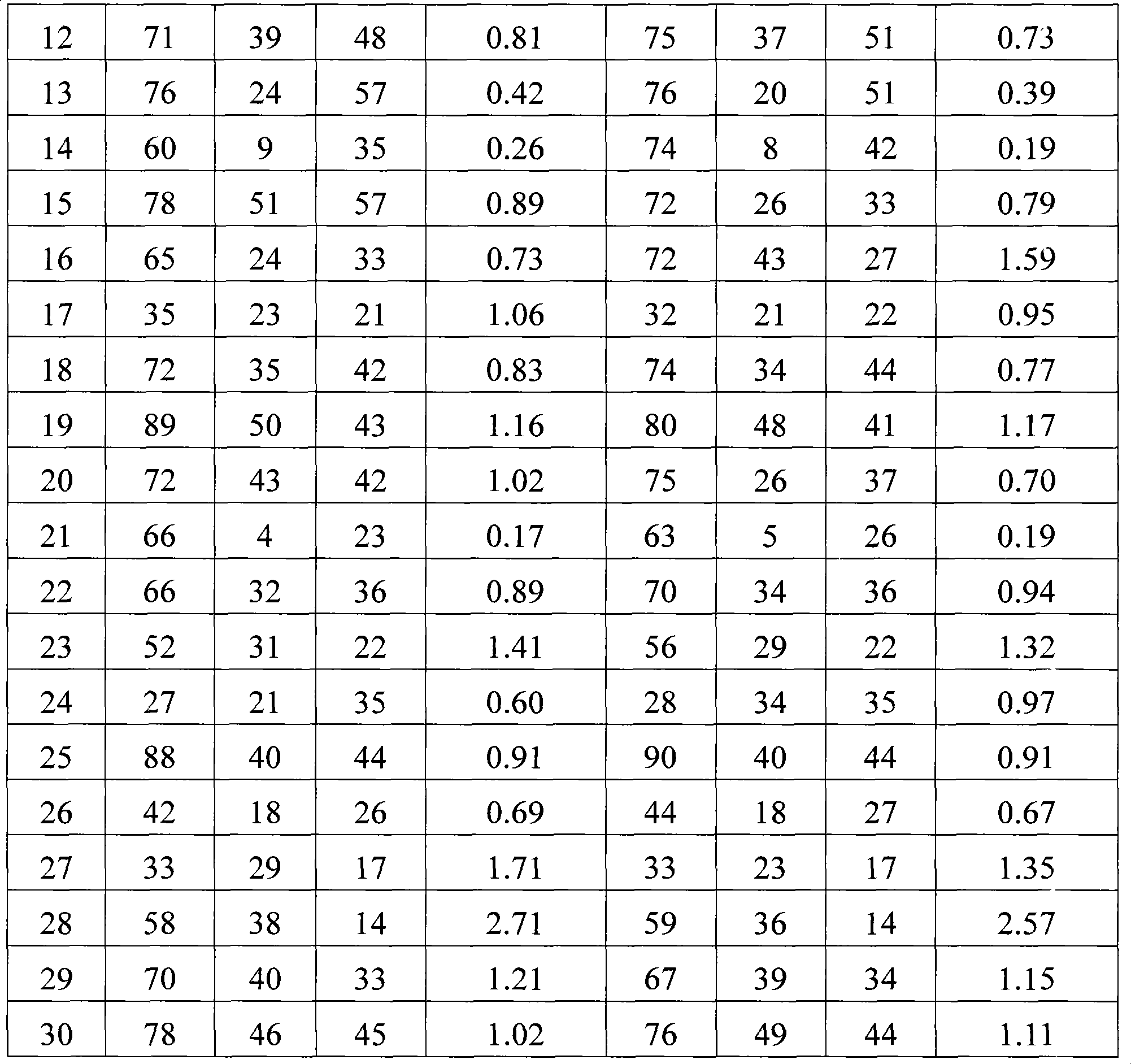
[0005] Although the lyophilized antibody-sensitized erythrocyte rosette reagent used in the McAb-A-E direct method itself has the characteristics of strong specificity, sensitivity, good stability, long validity period, convenient use, and can be detected only by an ordinary optical microscope, and the experimental results It can be stored for about two weeks and has been widely used in clinical laboratories and related research units for more than ten years. However, as a clinical detection method, the McAb-A-E direct method has many detection steps, relatively large sample volume, long cycle, It is easy to cause damage and loss of the tested cells, there is no quantitative control index for some important steps, and it is difficult to identify non-specific rosettes, which affects the counting results and other defects. It cannot meet the rapid and accurate clinical requirements, and many laboratories, especially clinical laboratories, accept Difficulty, which limits the generalization and application of this method

Method used


Image

  • Method for detecting lymphocyte subgroup with mono-clone antibody SPA hematid rosette method
  • Method for detecting lymphocyte subgroup with mono-clone antibody SPA hematid rosette method

Examples


Embodiment 1

[0021] Example 1 Lyophilized antibody sensitized erythrocyte rosette reagent WuT 3 、WuT 4 、WuT 8 (The freeze-dried product is a small amount of solid crystals, which dissolve after adding liquid, without affecting the volume). Add 0.5mL of calcium-magnesium-free Hank'S solution with a pH value of 7.0 to dissolve into a sensitized erythrocyte preservation solution. If the dissolved cell suspension cannot be used up, add 0.1% NaN 3 , sealed and stored at 4-6°C for 2-4 weeks.

[0022] Anticoagulate with heparin, extract 1mL of fasting venous blood from the patient, add 1mL of calcium- and magnesium-free Hank'S solution with a pH value of 7.0, mix well, and slowly add it to 2mL of lymphocyte separation medium with a dropper. Centrifuge at 800 rpm for 20 min in a 22 cm centrifuge.

[0023]Use a dropper to suck out the mononuclear cells and put them into another centrifuge tube with 4 to 6 times the amount of calcium- and magnesium-free Hank'S solution with a pH value of 7.0. As...

Embodiment 2

[0029] Example 2 Lyophilized antibody sensitized erythrocyte rosette reagent WuT 3 、WuT 4 、WuT 8 Add 0.5mL calcium-free and magnesium-free Hank'S solution with a pH value of 7.2 to dissolve into sensitized erythrocyte preservation solution. If the dissolved cell suspension cannot be used up, add 0.1% NaN 3 , sealed and stored at 4-6°C for 2-4 weeks.

[0030] Disodium salt of ethylenediaminetetraacetic acid (EDTA-Na 2 ) anticoagulant, draw 1.5mL of fasting venous blood from the patient, add 3mL of calcium- and magnesium-free Hank'S solution with a pH value of 7.2, mix well, and slowly add it to 2.5mL of lymphocyte separation solution with a dropper, and at 22°C, use A centrifuge with a radius of 22 cm was centrifuged at 1000 rpm for 18 min.

[0031] Aspirate the mononuclear cells with a dropper, put them into another centrifuge tube with 4 to 6 times the amount of calcium- and magnesium-free Hank'S solution with a pH value of 7.2, and suck as little lymphocyte separation s...

Embodiment 3

[0037] Example 3 Lyophilized antibody sensitized erythrocyte rosette reagent WuT 3 、WuT 4 、WuT 8 Add 0.5mL calcium-free and magnesium-free Hank'S solution with a pH value of 7.4 to dissolve into the sensitized red blood cell preservation solution. If the dissolved cell suspension cannot be used up, add 0.1% NaN 3 , sealed and stored at 4-6°C for 2-4 weeks.

[0038] Anticoagulate with heparin, extract 2 mL of fasting venous blood from the patient, add 4 mL of calcium- and magnesium-free Hank'S solution with a pH value of 7.4, mix well, and slowly add it to 3 mL of lymphocyte separation liquid with a dropper. Centrifuge at 1200rpm for 25min in a 22cm centrifuge.

[0039] Aspirate the mononuclear cells with a dropper, put them into another centrifuge tube with 4 to 6 times the amount of calcium- and magnesium-free Hank'S solution with a pH value of 7.4, and suck as little lymphocyte separation solution as possible. Mix well, centrifuge at 600 rpm, centrifuge for 6 minutes, d...


PUM


Abstract

The invention relates to a method for inspecting lymphocytic subgroup by a single clone antibody SPA red cell garland way. The method comprises the following procedures: first the red cell garland reagent is solved, then the circumference blood leukomonocytes are separated; leukomonocyte suspension is prepared by a Henke balancing salt solution free of Ca and Mg containing 20% serum of a newly borne calf; meanwhile, the sensitized red cell suspension is made into a sensitized red cell application liquid by using a Henke balancing salt solution free of Ca and Mg containing 20% serum of a newlyborne calf; then the leukomonocyte suspension and the sensitized red cell application liquid are mixed by 1:1 V / V into a mixed suspension, which is centrifuged after placing still under 18-28 centigrade, then is placed still for 15-45 min under 4 centigrade; finally the mixed suspension is absorbed 15 or 20 times by a pipet having a sucking head, the absorbed suspension is made into cell smear, which is naturally dried, dyed and counted by a microscope with high magnification. Comparing with prior McAb-A-E method, the invention is of simplified detection process, shorter period, quantitative controlling, and can meet clinic detection.

Description

technical field [0001] The invention relates to medical immunology, in particular to a method for detecting lymphocyte subsets by a monoclonal antibody SPA erythrocyte rosette method. Background technique [0002] With the development of molecular immunology, T, B, and NK lymphocytes can be divided into several subgroups according to the different clusters of differentiation (CD) on the surface of lymphocyte membranes. Clinical laboratories use anti-differentiation antigens CD3, CD4, CD8 , CD19, CD20, CD22, CD56, CD16 and other monoclonal antibodies (McAb), detect peripheral blood lymphocytes, and judge total T cells, T helper cells (TH), T suppressor / killer cells according to their positive rate (T s / T c ) subsets, B lymphocytes, NK cells, etc., to judge the immune function of the human body, to achieve preventive health care and disease diagnosis, treatment, and prognosis (according to the test results, the doctor predicts the future development of a disease) Purpose. ...

Claims


Application Information

Patent Timeline
Patent Type & Authority Patents(China)
IPC IPC(8): G01N33/577G01N21/25C12Q1/02
Inventor 姚伯程
Owner 甘肃省医学科学研究院
Learn More