
Method and device for producing shaped microbial cellulose for use as biomaterial especially for microsurgery

A technology for microbial cellulose and cellulose generation, applied in the field of preparing shaped microbial cellulose

Inactive Publication Date: 2009-01-14
SURA CHEM GMBH
View PDF7 Cites 4 Cited by
  • Summary
  • Abstract
  • Description
  • Claims
  • Application Information

AI Technical Summary

Problems solved by technology

However, use in small vessels (2-3 mm in diameter in the cases described) is of concern due to the risk of thrombotic deposition

Method used


Image

  • Method and device for producing shaped microbial cellulose for use as biomaterial especially for microsurgery
  • Method and device for producing shaped microbial cellulose for use as biomaterial especially for microsurgery

Examples


Embodiment Construction

[0025] The culture broth is sterilized in a known manner, inoculated with cellulose-producing bacteria, such as the microorganism Acetobacter xylinum (xylanacetobacter), which produces a stable cellulose layer, and between the module walls, for example at 28° C. to 30° C. Cultivate in the gap. The biological material (cellulose) produced during the cultivation is separated from the walls of the modules and purified (see EP 396 344 A3).

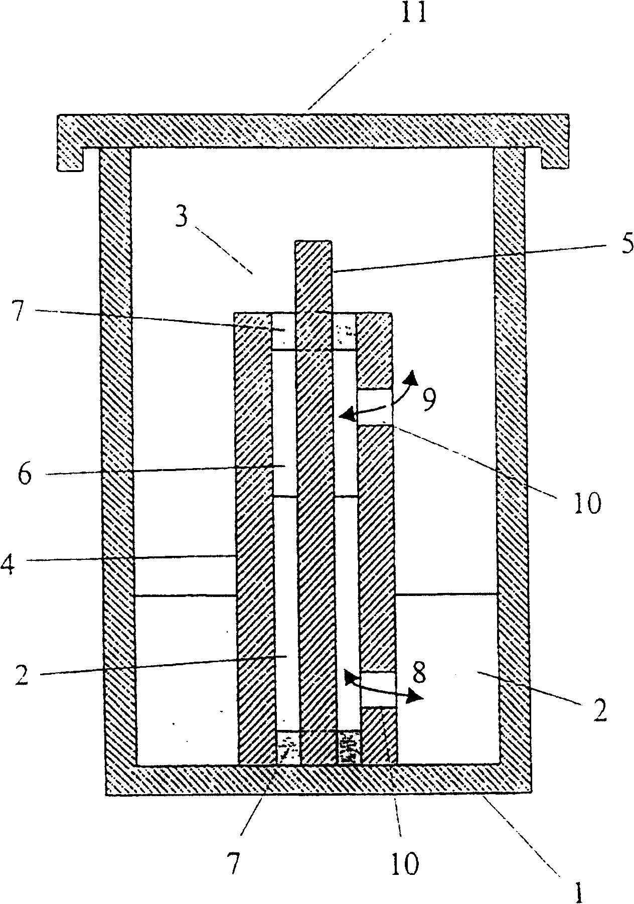
[0026] According to the invention, however, the inoculated culture solution is not injected into the spaces between the walls of the modules, for example between glass master molds (Glasmatrix) which are preferably made of glass parts which can be separated from each other, but the molds are removed during the cultivation process. The part wall (glass master mold) is immersed in the container containing the inoculated culture solution, so that the culture solution enters the space between the module walls by capillary force. This ensures a mo...


PUM

PropertyMeasurementUnit
diameteraaaaaaaaaa
diameteraaaaaaaaaa
thicknessaaaaaaaaaa

Abstract

The use of exogenic materials for replacing blood vessels carries the risk of thrombosis and is therefore particularly unsuitable for microsurgical applications (inner vessel diameters of 1-3 mm and less), or only suitable under certain conditions. Replacements of blood vessels with a very small lumen in particular require biomaterials which guarantee that the surfaces of the prosthesis that come into contact with the blood are of a very high quality, and which reliably avoid this kind of thrombosis adhesion. The biomaterial is produced by immersing shaped body walls, especially of a glass matrix consisting of a glass tube and glass body, in a container of an inoculated nutrient solution so that the inoculated nutrient solution is drawn into the area between the walls of the shaped body and cultivation takes place in a moist, aerobic environment. In each subsequent cultivation process, an unused shaped body (glass body) is used as the shaped body wall for shaping the surface of the prosthesis material that is to come into contact with the blood when the biomaterial is used. This is the only sure way of reproducing the high surface quality of the vessel prosthesis and hereby reliably preventing thrombosis adhesion on the biomaterial used. The inventive method is particularly suitable for microsurgical applications, especially for replacing blood vessels and other internal hollow organs or as a cuff for covering nerve fibres, etc.

Description

technical field [0001] The present invention relates to a method and a device for the preparation of shaped microbial cellulose which can be used as a biomaterial, in particular as a biomaterial in microplastic surgery, e.g. for angiogenesis and other The internal hollow organs or the rings used to wrap nerve fibers, etc. Background technique [0002] It is known (e.g. JP 3 165 774 A1) that microbial cellulose can be used as a biomaterial for plastic surgery, e.g. for tissue transplantation of the abdominal wall, skin, subcutaneous tissue, organs, digestive tract, esophagus, respiratory tract and urinary tract as well as cartilage and adipose tissue . [0003] It is also known (for example JP 8 126 697 A2, EP 186 495 A2, JP 63 205 109 A1, JP3 165 774 A1) that microbial cellulose can be made into special application shapes during the preparation process, such as sheet, rod, cylinder shape and strip etc. [0004] The following preparation methods have been described: [00...

Claims


Application Information

Patent Timeline
Patent Type & Authority Patents(China)
IPC IPC(8): C12P19/04A61L17/00A61L27/20A61L29/04C08L1/02C12R1/02C12M1/00A61L31/00
CPCC12P19/04C08L1/02
Inventor 迪特尔·克莱姆西尔维亚·马尔施迪特尔·舒曼乌尔丽克·乌德哈尔特
Owner SURA CHEM GMBH
Learn More