
Amniotic membrane covering for a tissue surface and devices facilitating fastening of membranes

A technology for surface coverings and coverings, which can be used in applications, eye implants, and preservation of human or animal bodies, and can solve the problem of time-consuming cultivation and nesting

Inactive Publication Date: 2007-12-12
TISSUETECH INC
View PDF6 Cites 2 Cited by
  • Summary
  • Abstract
  • Description
  • Claims
  • Application Information

AI Technical Summary

Problems solved by technology

But making culture nests by sewing is time consuming

Method used


Image

  • Amniotic membrane covering for a tissue surface and devices facilitating fastening of membranes
  • Amniotic membrane covering for a tissue surface and devices facilitating fastening of membranes
  • Amniotic membrane covering for a tissue surface and devices facilitating fastening of membranes

Examples


Embodiment approach

[0210] Prototype 1: Immobilization of amniotic membranes on culture nests

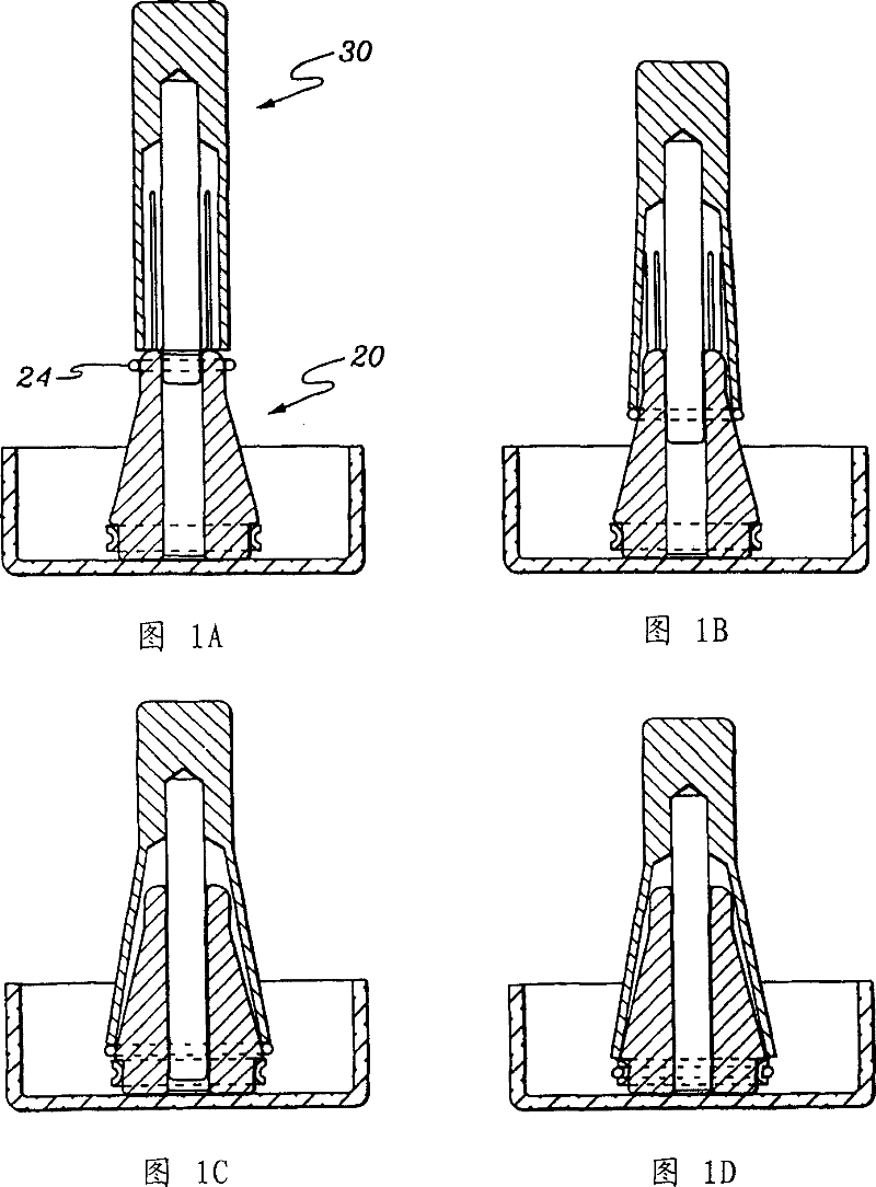
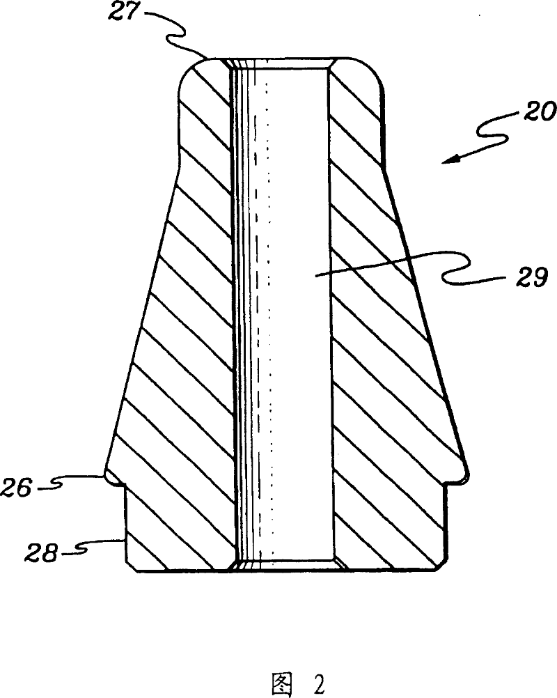
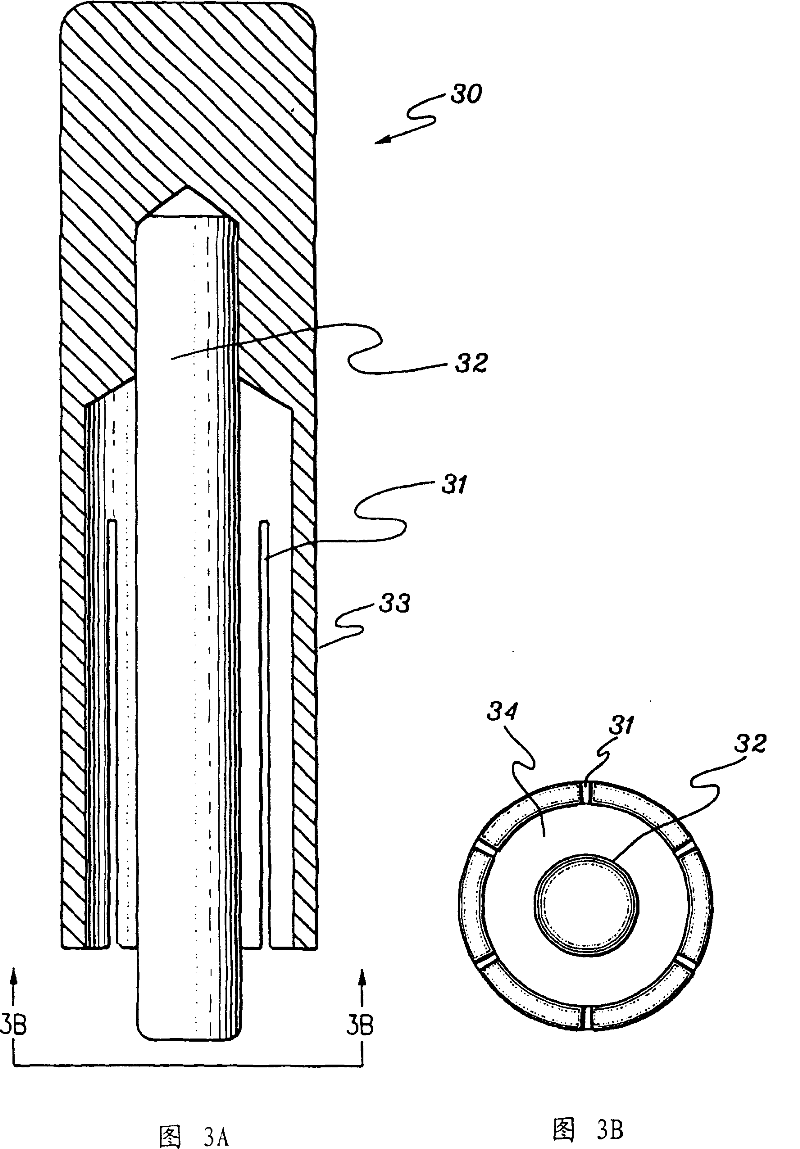
[0211] Prototype 1 consisted of a set of devices shown in Figures 1A-1D and Figures 2, 3A and 3B. These devices include a conical dilator as shown in Figures 1A-1D (20) and Figure 2; a device (30) for frictionally engaging an elastic ring (24), and a tool (30) for transferring said elastic ring to the nest. (as shown in Figures 1A-1D, the ring on the base of the dilator (20)). The original purpose of these devices was to assist in the fixation of the amnion on the culture nest, i.e. the culture of cells on the amnion base. Thus, Prototype 1 illustrates how the amniotic membrane can be fixed to a solid support, the culture nest, using elastic loops. The operation method of making prototype 1 with these devices includes the following steps shown in Fig. 1A-1D.

[0212] Step 1: Place the elastic ring (24) on the apex of the conical dilator (20) with the base of the dilator in contact with the ring whic...


PUM

PropertyMeasurementUnit
weightaaaaaaaaaa
hardnessaaaaaaaaaa

Abstract

The present invention relates to a biopolymer covering for a tissue surface including, for example, a dressing, a bandage, a drape such as a bandage contact lens, a composition or covering to protect tissue, a covering to prevent adhesions, to exclude bacteria, to inhibit bacterial activity, or to promote healing or growth of tissue. An example of such a composition is an amniotic membrane covering for an ocular surface. Use of a covering for a tissue surface according to the invention eliminates the need for suturing. The invention also includes devices facilitating the fastening of a membrane to a support, culture inserts, compositions, methods, and kits for making and using coverings for a tissue surface and culture inserts. Compositions according to the invention may include cells grown on a membrane or attached to a membrane, and such compositions may be used as scaffolds for tissue engineering or tissue grafts. A method of preparing and using an amniotic membrane covering for a tissue surface as a controlled release drug delivery vehicle is also disclosed.

Description

[0001] related application [0002] This application claims the benefit of US provisional application 60 / 365,356, filed March 14, 2002, the teachings of which are incorporated by reference in their entirety. [0003] governmental support [0004] The present invention is fully or partly sponsored by a research grant (No. EY06819) from the National Institutes of Health of the United States, and the US government has partial rights to the present invention. Background technique [0005] Amnion compositions, such as amnion extracts and amnion obtained from mammalian amniotic tissues such as humans, pigs, or horses, contain biological growth factors. Amnion is a biological membrane that lines the inner wall of the amniotic cavity and consists of a simple cuboidal epithelium, a thick basement membrane, and an avascular mesenchymal layer containing hyaluronic acid. Amnion compositions are known to be anti-inflammatory and reduce fibrovascular ingrowth and promote epithelializat...

Claims


Application Information

Patent Timeline
Patent Type & Authority Patents(China)
IPC IPC(8): A01N1/02A61F2/14A61F9/007A61F9/00A61F9/04A61L27/00A61L27/36A61L27/38A61L31/00
CPCA61L31/005A61L27/3679A61L27/3813A61L27/3882A61L27/3604A61F9/0017A61F9/04
Inventor 谢弗·C·G·曾赫尔加·桑多瓦尔威廉·G·李
Owner TISSUETECH INC
Learn More