
Device and method for treating retinal detachment

Active Publication Date: 2018-02-20
TARABISHY AHMAD BAKIR
View PDF3 Cites 2 Cited by
  • Summary
  • Abstract
  • Description
  • Claims
  • Application Information

AI Technical Summary

Benefits of technology

The proposed device helps to accurately place a drainage tube in the retina and allows for controlled suction drainage of subretinal fluid. It allows for careful and controlled drainage without causing unnecessary trauma to the retina and avoids the need for an extensive dissection or creating iatrogenic retinal breaks.

Problems solved by technology

Retinal detachment is a common cause of severe vision loss.
A tear typically develops when there is a sudden collapse of the vitreous thus causing tugging on the retina with enough force to cause a tear.
In areas in which the retina is detached and the cells lack oxygen, vision may be lost if not treated quickly.
However, these techniques have significant disadvantages and complications.
For example, external drainage with a sclerotomy requires an extensive dissection and cannot be done while directly visualizing the retina.
External sclerotomy can also be complicated by hemorrhage, retinal incarceration, or iatrogenic retinal damage.
Given the disadvantages in the current techniques for treating retinal detachment, what is needed is a device and system which would allow drainage site selection, drain placement and subretinal fluid drainage under direct visualization using an internal wide angle viewing system.

Method used


Image

  • Device and method for treating retinal detachment
  • Device and method for treating retinal detachment
  • Device and method for treating retinal detachment

Examples


Embodiment Construction

[0029]In the following detailed description of the preferred embodiments, reference is made to the accompanying drawings, which form a part hereof, and within which are shown by way of illustration specific embodiments by which the invention may be practiced. It is to be understood that there are other embodiments by which the invention may be practiced. It is to be understood that other embodiments may be utilized and structural changes may be made without departing from the scope of the invention.

Definitions

[0030]Where a range of values is provided, it is understood that each intervening value, to the tenth of the unit of the lower limit, unless the context clearly dictates otherwise, between the upper and lower limits of that range is also specifically disclosed. Each smaller range between any stated value or intervening value in a stated range and any other stated or intervening value in that stated range is encompassed in the invention. The upper and lower limits of these small...


PUM


Abstract

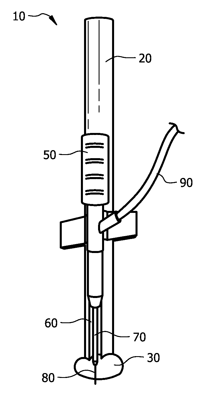
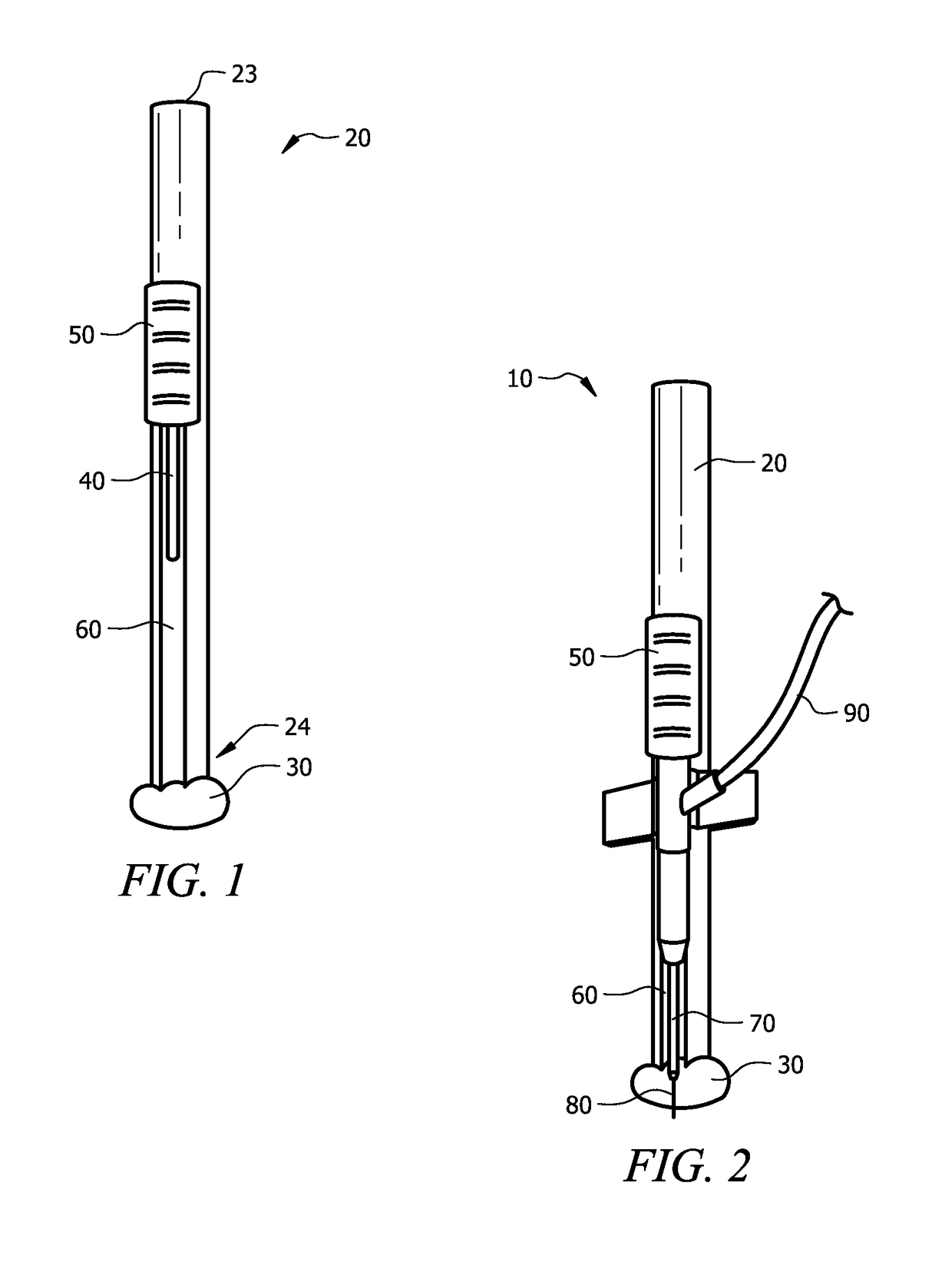
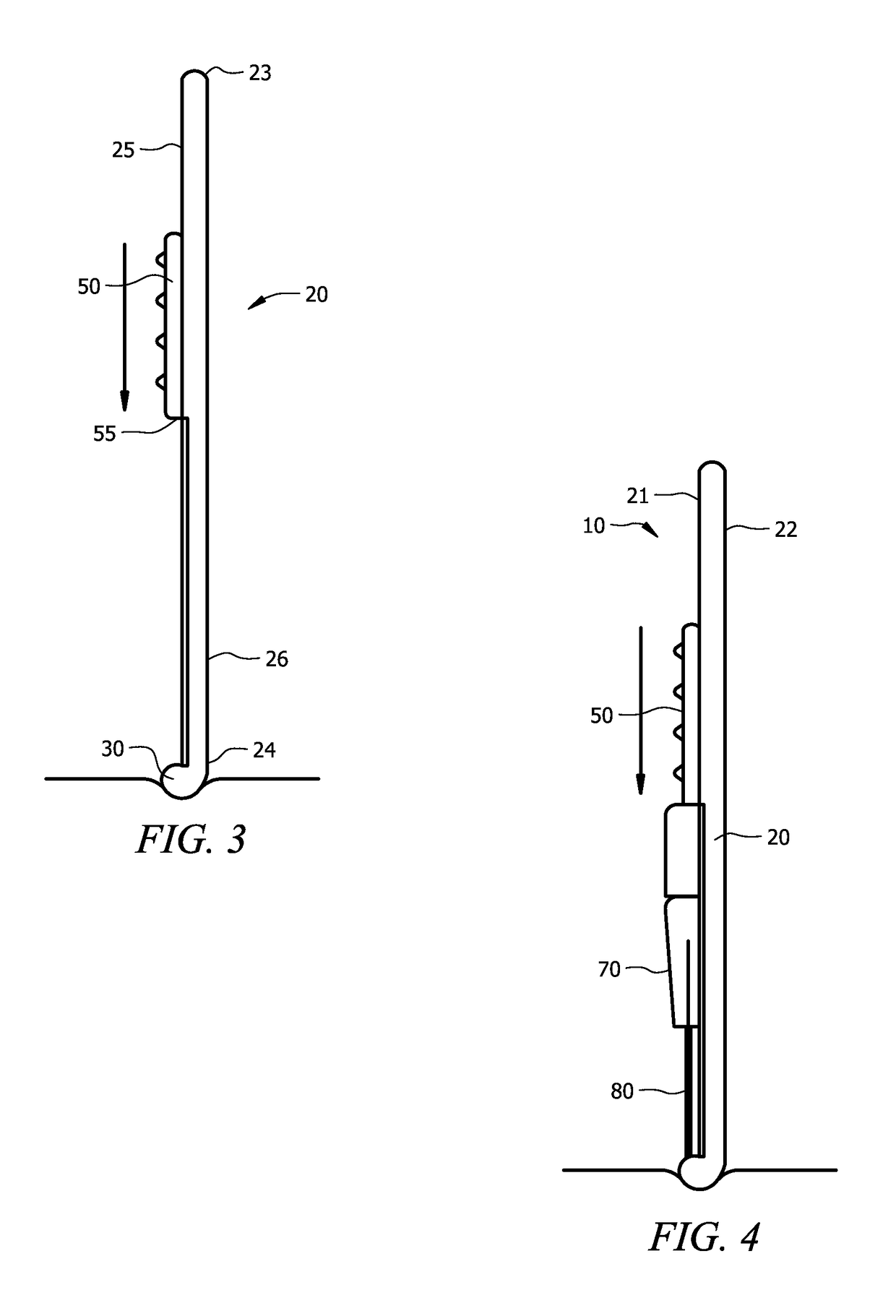
A device, system and method for treating retinal detachment is presented. The device is generally comprised of a depressor, a cannula and a needle. In use, the depressor is used to create an indentation on the surface of the eye that can be viewed internally to determine the site for drainage. A cannula advancement mechanism is engaged and the cannula containing the needle is inserted into the determined drainage site. The needle is removed and an aspiration device is connected to the cannula to drain the subretinal fluid to treat retinal detachment.

Description

CROSS REFERENCE TO RELATED APPLICATION[0001]This application is a nonprovisional of and claims priority to U.S. Provisional Patent Application No. 62 / 020,595, entitled “CANNULATED NEEDLE INTRODUCER TO DRAIN SUBRETINAL FLUID FOR TREATING RETINAL DETACHMENT,” filed on Jul. 3, 2014 by the same inventor, the contents of which are hereby incorporated by reference.FIELD OF THE INVENTION[0002]This invention relates to a novel device and method for treating retinal detachment. More particularly, it relates to a device and method that allows direct visualization of the retina for drain placement and subretinal fluid drainage.BACKGROUND OF THE INVENTION[0003]Retinal detachment is a common cause of severe vision loss. In retinal detachment, the retina pulls away from the layer of blood cells which provide oxygen thus leaving the retinal cells lacking oxygen. Retinal detachment can occur when vitreous fluid leaks through a retinal hole or tear and collects underneath the retina. Tears or holes ...

Claims


Application Information

Patent Timeline
IPC IPC(8): A61M5/00A61M1/00A61M27/00A61F9/007
CPCA61F9/00736A61F9/00727A61M27/00A61M1/008
Inventor TARABISHY, AHMAD BAKIR
Owner TARABISHY AHMAD BAKIR
Learn More