
Interventional imaging system

a technology of imaging system and interventional body, which is applied in the field of interventional medical diagnosis and/or therapy system, can solve the problems of unsuitability of access path, limited body volume, and inability of physicians to identify, so as to achieve low calibration and registration effort

Active Publication Date: 2018-02-13
SIEMENS HEALTHCARE GMBH
View PDF8 Cites 0 Cited by
  • Summary
  • Abstract
  • Description
  • Claims
  • Application Information

AI Technical Summary

Benefits of technology

[0016]The object of the invention is to provide an interventional imaging system and method which allow for an intervention to be conducted in accordance with an intervention plan, to be supported and monitored by ongoing imaging, in particular radioscopy, with which at the same time the effort in calibration and registration can be kept low, and which functions without an additional location system.
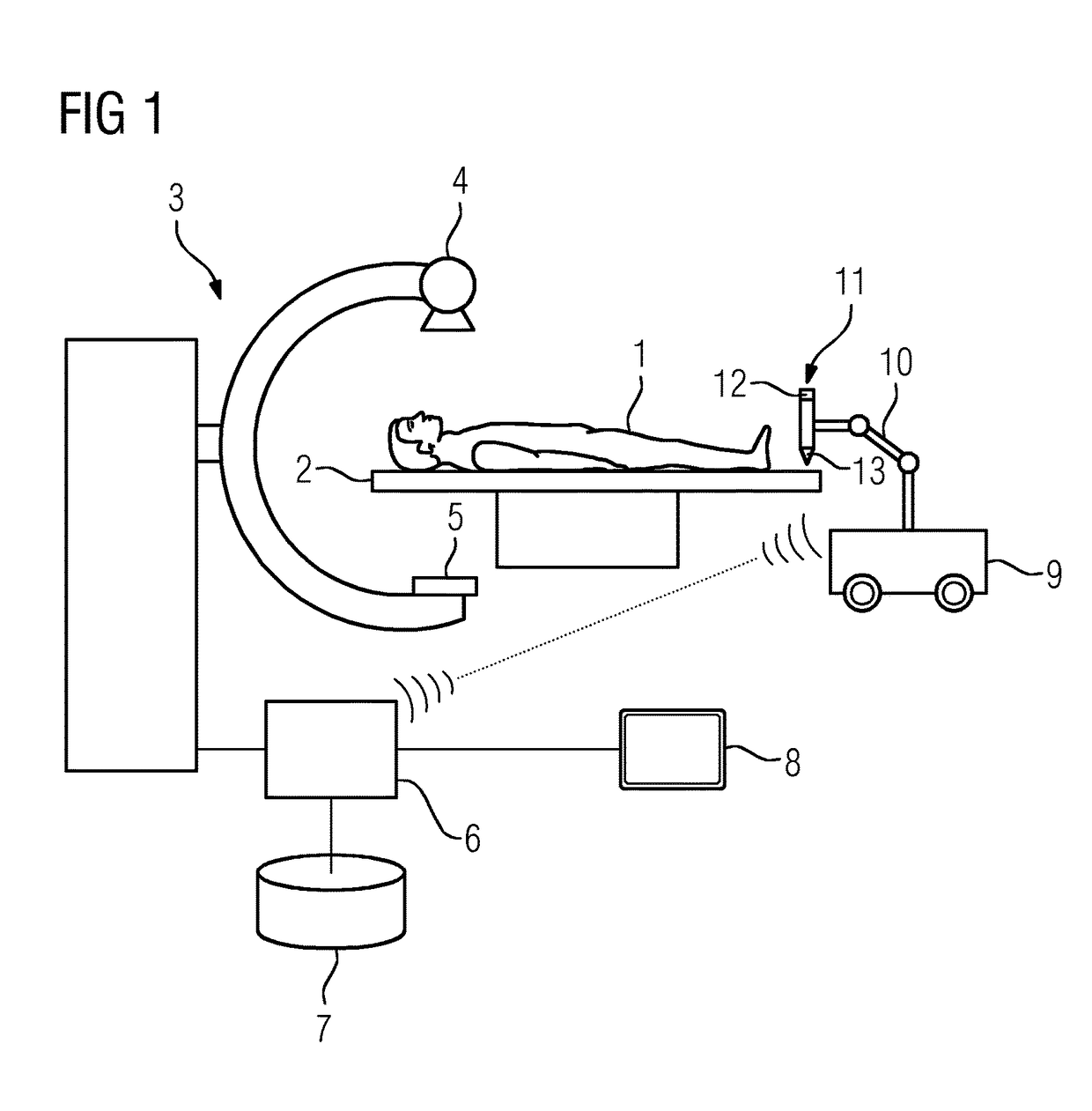
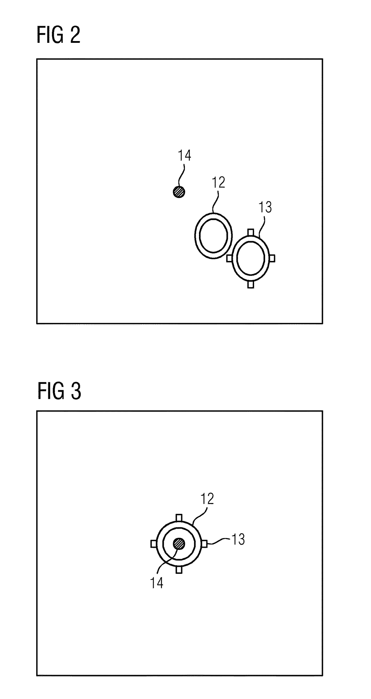
[0040]A basic concept of the invention in accordance with the foregoing explanations accordingly consists of the use of continuous interventional imaging (radioscopy) for the final and precise positioning of the instrument holding or guidance of an intervention instrument in accordance with a predetermined planning. As the imaging system, use is made preferentially of an X-ray system, e.g. a C-arm X-ray system. Accordingly, a system and method are described which allow for simple manual pre-positioning, which can be easily moved to and from the patient couch, function without previous calibration, registration, or a location system, and attain an extremely high degree of flexibility.
[0046]The imaging itself can be carried out with an adjusted low image rate. Because the markers can be optimally recorded, i.e. impervious to X-rays in the case of an X-ray system, the fluoroscopic dose per image can also be substantially reduced.

Problems solved by technology

Due to the fact that the physician cannot directly see the instrument in the body of the patient, he is reliant on the support of imaging methods.
Problems arise, for example, in situations involving adipose or heavily-built patients.
Due to the technical realization of the C-arm X-ray device, only a limited volume of the body can be reconstructed.
With virtual planning, in this situation the physician cannot identify where the insertion point is located, and whether the planned access path may possibly be unsuitable for the intervention due to the superimposition of bones or ribs.
This procedure, as well as involving unpleasant effects for the patient, also incurs an increased risk of complications, such as bleeding, organ injury, or hematoma.
In addition to this, the precision of the puncture is restricted with this procedure, in particular with very small target regions.
This means, however, that account cannot be taken of deformations of the instrument, e.g. of the tip of a needle, caused due to resistances in the body, and which under certain circumstances may lead to a deviation from the puncture path.
If such a system is to be used in the surgical environment, however, a number of limitations may arise.
This calibration is prone to faults, since random mechanical effects on the instrument holder in the operating theater cannot be excluded.
This is time-consuming and not practicable in the operation environment.
The large number of registration steps is elaborate and time-consuming.
In addition to this, additional location systems must be omitted, since their hardware is often in the way, as well as prone to faults.

Method used


Image

  • Interventional imaging system
  • Interventional imaging system
  • Interventional imaging system

Examples


Embodiment Construction

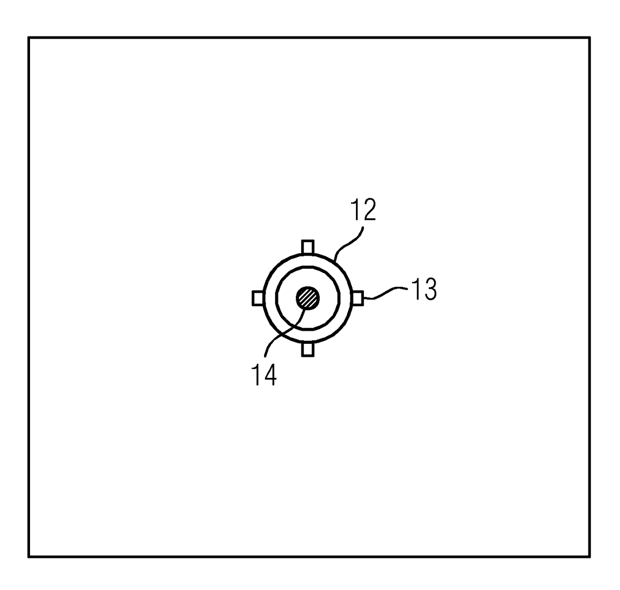
[0054]Shown in diagrammatic form in FIG. 1 is an interventional imaging system. This comprises a C-arm X-ray system 3, a patient couch 2, and a cart 9 equipped for intervention purposes.

[0055]The C-arm X-ray system 3 comprises an X-ray emitter 4 and an image detector 5 arranged on a C-arm. It is connected to a navigation facility 6, which serves to support interventions which take place with the aid of interventional image data acquired by the C-arm X-ray system 3. According to a simple embodiment, intervention data is acquired in 2D by the C-arm X-ray system 3. In a more elaborate embodiment, however, 3D data can also be acquired, for which purpose the detector normals, which are derived from the position of the C-arm, must be rotated.

[0056]In order to carry out an intervention, the navigation facility 6 loads pre-intervention data from a corresponding data memory 7. The pre-intervention data is, as a rule, 3D data, which is acquired from a body which is to be treated before an int...


PUM


Abstract

An interventional medical diagnosis and / or therapy system is provided. The system provides an interventional imaging system and method which allows for an intervention, to be conducted in accordance with an intervention plan, to be supported and monitored by ongoing imaging, in particular radioscopy, with which, at the same time the effort in calibration and registration is kept low, and which functions without an additional location system. The interventional imaging system includes an imaging device to record intervention data of a body, at least two position markings, capable of being recorded with the imaging device, for the marking of an intervention instrument, a display apparatus to reproduce recorded intervention data and position markings, a navigation facility connected to the display apparatus to load pre-intervention data of the body, in which an intervention location of the body is contained, and for the mutual registration of the pre-intervention data with the intervention data.

Description

CROSS REFERENCE TO RELATED APPLICATIONS[0001]The present application claims priority to German application No. 102013213727.3 DE filed Jul. 12, 2013, the entire content of which is hereby incorporated herein by reference.FIELD OF INVENTION[0002]The invention relates to an interventional medical diagnosis and / or therapy system.BACKGROUND OF INVENTION[0003]Interventional procedures, such as are applied, for example, in radiology, are today already playing an important part in the diagnosis and therapy of many illnesses. By way of example, certain instruments are used, such as needles, to reach a specific point in the body or inside an organ from the outside through the skin (percutaneous). This relates for instance to punctures, biopsies, ablations, or brachytherapies, or fixations, such as screws, are put in place.[0004]Based on further technical developments of the imaging systems, the techniques referred to as modalities, such as, for example, computed tomography, magnetic resonanc...

Claims


Application Information

Patent Timeline
Patent Type & Authority Patents(United States)
IPC IPC(8): A61B34/30A61B6/12A61B6/00A61B34/20A61B90/00
CPCA61B6/12A61B6/486A61B6/5217A61B6/488A61B2090/3966A61B34/20A61B34/30A61B2090/376A61B6/5247
Inventor HARTKENS, THOMASREDEL, THOMAS
Owner SIEMENS HEALTHCARE GMBH
Learn More