
pH-sensitive fluorescent sensors for biological amines

a fluorescent sensor and biological amine technology, applied in biological testing, material testing goods, organic chemistry, etc., can solve the problems of lack of spatial resolution, poor throughput, and limited non-optical techniques

Active Publication Date: 2018-01-09
UNIVERSITY OF MISSOURI
View PDF2 Cites 0 Cited by
  • Summary
  • Abstract
  • Description
  • Claims
  • Application Information

AI Technical Summary

Benefits of technology

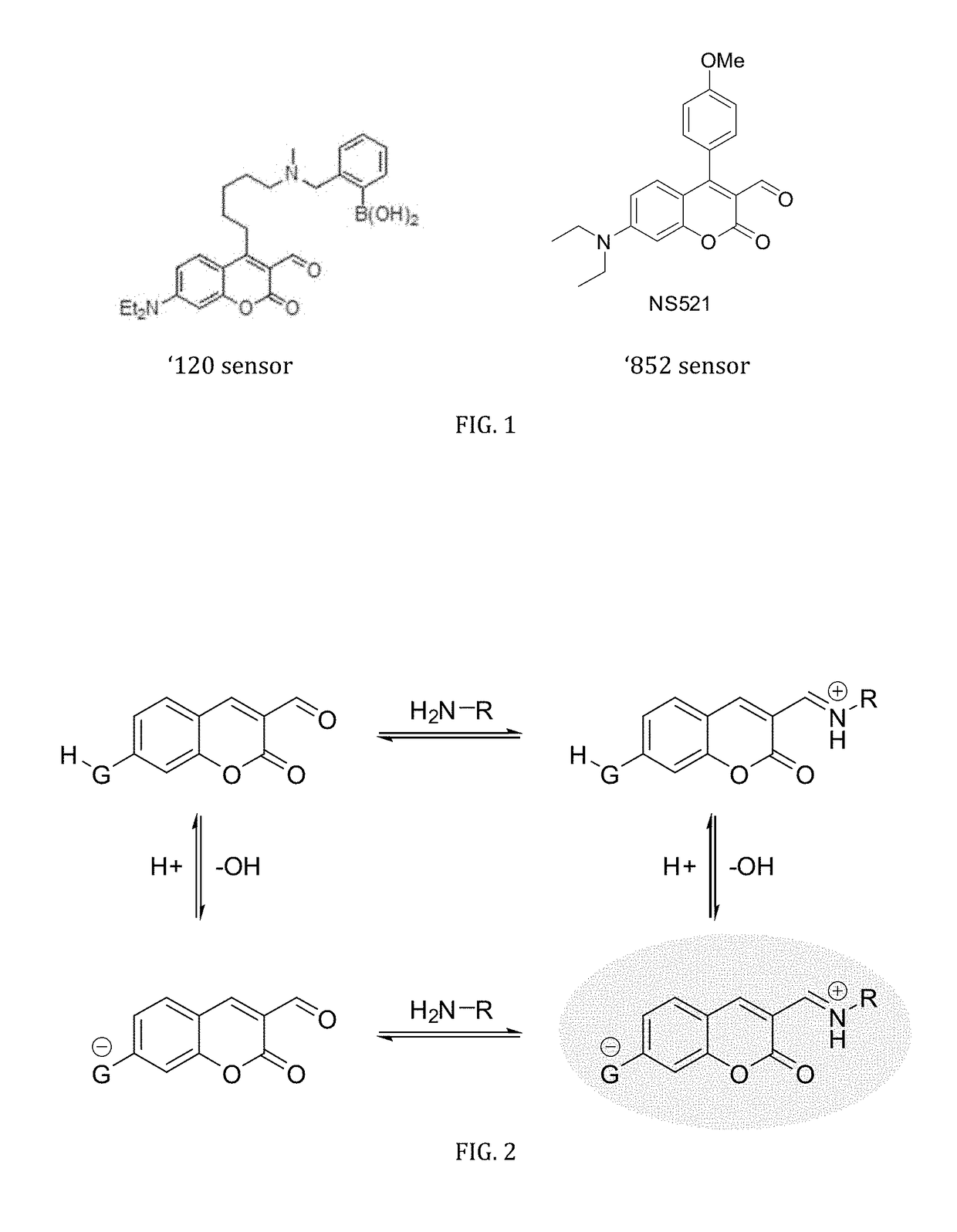
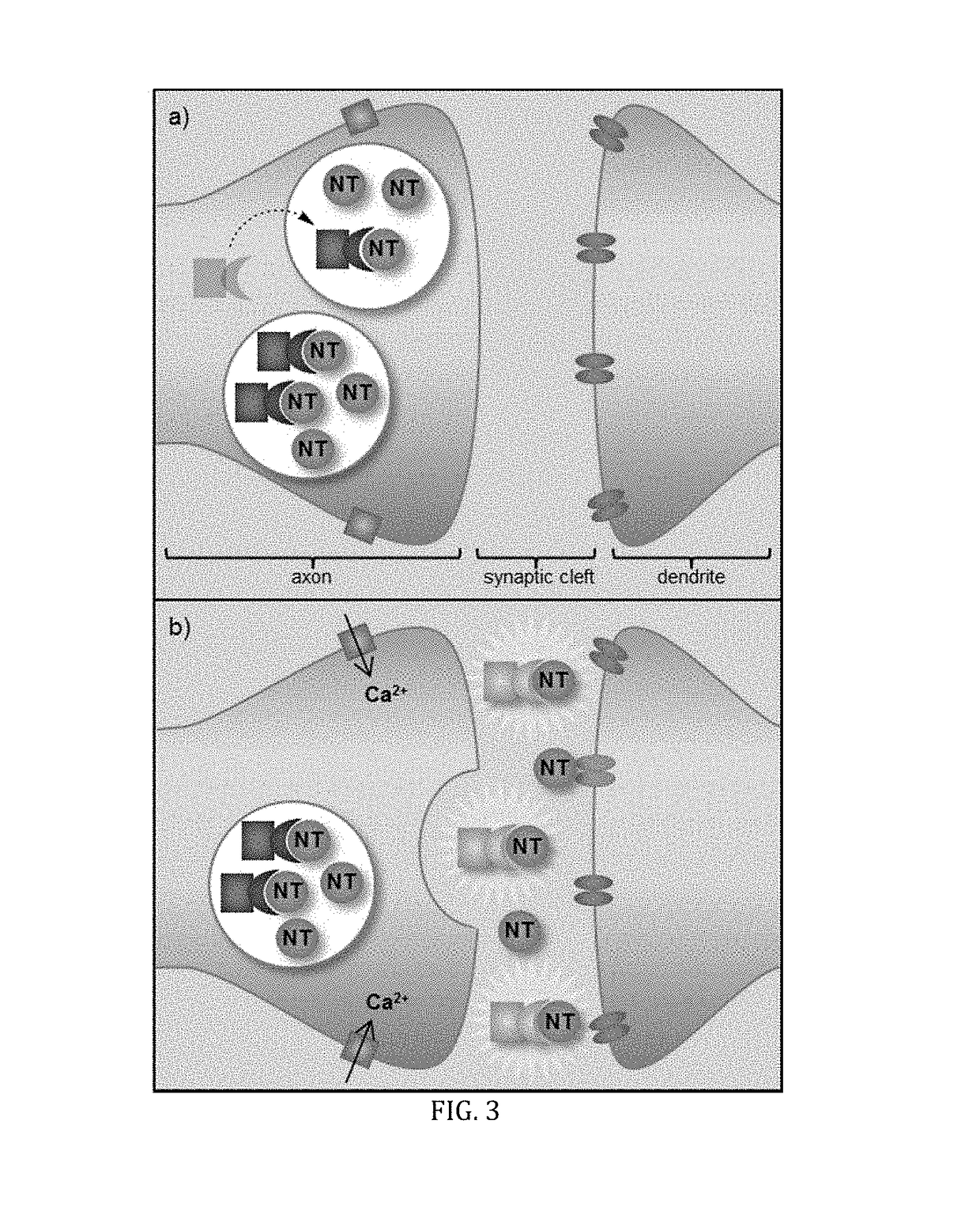
The present invention provides a series of pH-sensitive fluorescent sensors for imaging exocytosis of the neurotransmitters. These sensors selectively label primary amine neurotransmitters found at high concentrations within vesicles and concomitantly allow for direct visualization of only active neurotransmitters released upon exocytosis by capitalizing on the pH gradient between the vesicle and the synaptic cleft. The sensors have a pKa in a range of about 6.0 to about 7.4. The invention also includes a method of detecting one or more primary neurotransmitters released during exocytosis.

Problems solved by technology

Non-optical techniques are limited by poor throughput and a lack of spatial resolution.
These methods solely monitor the process of vesicle membrane fusion during an exocytotic event but do not directly image active neurotransmitters released upon exocytosis.
However, these protein-based biosensors require genetic manipulation and display high, irreversible affinity for glutamate with limited dynamic range and overall small changes in fluorescence.
As a result, these protein-based sensors are neither intended for, nor compatible with, specialized neurosecretory cells (e.g., glutamatergic, dopaminergic, etc.) that possess high concentrations of primary amine neurotransmitters (300 mM-1M).
There remains, however, a scarcity of small molecular sensors that can directly detect and image neurotransmitters upon exocytosis.

Method used


Image

  • pH-sensitive fluorescent sensors for biological amines
  • pH-sensitive fluorescent sensors for biological amines
  • pH-sensitive fluorescent sensors for biological amines

Examples


example 1

of ES517

[0058]Compound 1 (465 mg, 1.746 mmol) and N-phenyltriflimide (686 mg, 1.921 mmol) were combined in a round bottom flask. THF (24 mL) was added and then DIPEA was added dropwise (0.38 mL, 2.270 mmol). The mixture stirred at ambient temperature for 3 h followed by removal of the solvent in vacuo. The residue was purified by chromatography (95:5 CH2Cl2 / EtOAc) to yield Compound 2 (607.2 mg, 87%) as a golden oil: 1H NMR (500 MHz, CDCl3) δ 9.95 (s, 1H), 7.56-7.62 (m, 3H), 7.33-7.37 (m, 2H), 7.29-7.32 (m, 2H), 7.16 (dd, 1H, J=9.0, 2.5 Hz); 13C NMR (125 MHz, CDCl3) δ 187.5, 159.5, 157.1, 155.0, 152.6, 131.4, 130.8, 130.3, 129.0, 128.4, 119.9, 119.7 (q, C—F, J=40 Hz), 118.0, 117.3, 110.6; IR (neat, cm−1) 1765, 1605, 1552, 1422, 1364, 1217, 1136, 1107, 980; HRMS calculated for C17H9F3O6SNa (M+Na+): 420.9964. Found: 420.9961.

[0059]Compound 2 (123 mg, 0.309 mmol) was combined with N,N-dimethylsulfamide (42 mg, 0.340 mmol), Pd2dba3 (14 mg, 0.015 mmol), SPhos (18 mg, 0.046 mmol), and K3PO...

example 2

opic Property Studies of ES517

[0060]FIG. 6 shows results of initial binding studies performed by titrating ES517 with glutamate and exciting at 488 nm, a region that is sufficiently red to prohibit absorbance and subsequent emission from the unbound sensor. A 12-fold fluorescence enhancement was obtained upon binding to glutamate though with a low binding constant (Ka=8.6 M−1). While low, this binding constant is believed to be preferred for reversible binding due to the high millimolar concentration of glutamate in a vesicle (˜300 mM).

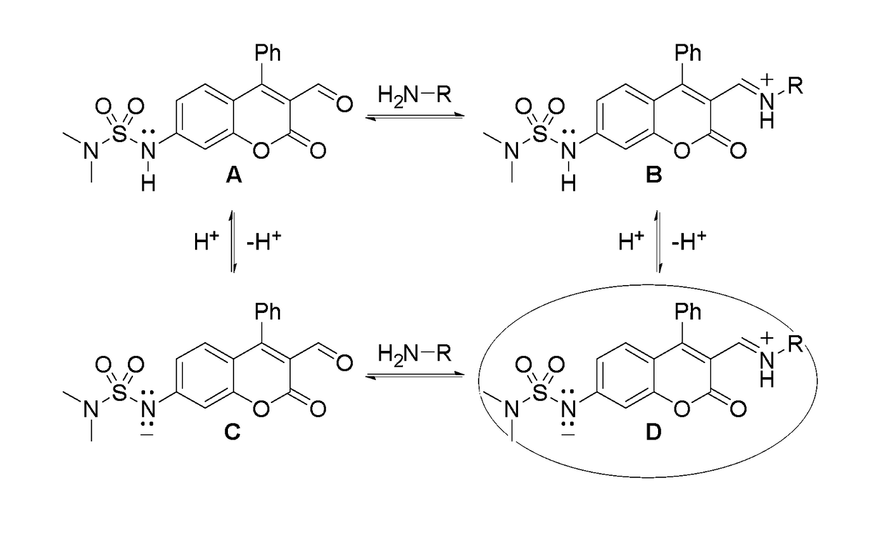
[0061]FIG. 7 shows the tests of the pKa of the bound sensor. ES517 was saturated with glutamate and the spectroscopic properties measured over a wide pH range. The protonated form of the bound sensor (FIG. 4, structure B) absorbed at 368 nm. Upon deprotonation, two bands were observed at 428 and 458 nm. The 428 nm band was assigned to the unbound deprotonated form of the sensor (structure C). The 458 nm band therefore represents the bound, deprotonate...


PUM

PropertyMeasurementUnit
pKaaaaaaaaaaa
pHaaaaaaaaaa
pKaaaaaaaaaaa

Abstract

The invention is directed to dual-analyte fluorescent chemosensors for the direct detection and visualization (imaging) of neurotransmitters released upon exocytosis. The inventive sensor exploits the high concentration of neurotransmitters (e.g., glutamate, norepinephrine, and dopamine) and capitalizes upon the pH gradient between the vesicle and synaptic cleft.

Description

CROSS-REFERENCE TO RELATED APPLICATIONS[0001]This application claims the benefit of provisional application, U.S. Ser. No. 61 / 958,610, filed Aug. 1, 2013, which is incorporated herein by reference in its entirety.STATEMENT REGARDING FEDERALLY SPONSORED RESEARCH OR DEVELOPMENT[0002]This invention was made with Government support under Grant No. CHE-1112194 awarded by the National Science Foundation. The Government has certain rights in the invention.FIELD OF THE INVENTION[0003]The present invention relates to fluorescent sensors for intracellular analyte detection. More particularly, the present invention relates to fluorescent sensors for detecting and imaging neurotransmitters upon exocytosis.BACKGROUND OF THE INVENTION[0004]Neurotransmitters are critical to the regulation of the central and peripheral nervous systems and command a number of functions such as learning, memory, sleep, and movement. Deng et al., Determination of amino acid neurotransmitters in human cerebrospinal flu...

Claims


Application Information

Patent Timeline
Patent Type & Authority Patents(United States)
IPC IPC(8): C07D311/16G01N33/74
CPCG01N33/74C07D311/16
Inventor GLASS, TIMOTHY EGILLIS, KEVIN DHETTIE, KENNETH SKLOCKOW, JESSICA L
Owner UNIVERSITY OF MISSOURI
Learn More