Cost-sensitive linear reconstruction based optic cup localization

a localization and optic cup technology, applied in the field of cost-sensitive linear reconstruction based optic cup localization, can solve the problems of inability to recover glaucoma vision loss, ineffective glaucoma screening in the whole population, loss of peripheral vision, etc., and achieve the effect of effective and efficien

Inactive Publication Date: 2017-06-13
AGENCY FOR SCI TECH & RES
View PDF12 Cites 0 Cited by
  • Summary
  • Abstract
  • Description
  • Claims
  • Application Information

AI Technical Summary

Benefits of technology

[0024]The present invention aims to provide an effective and efficient method to obtain, from one or more retinal images (particularly non-stereo images), data characterizing the optic cup, such as data indicating the location and / or size of the optic cup. The location and / or size may be expressed in relation to a pre-specified optic disc, such as an optic disc segmented by ASM or other established algorithms.

Problems solved by technology

Progression of the disease results in loss of peripheral vision, so patients may complain of “tunnel vision” (only being able to see the centre).
Vision loss in glaucoma cannot be recovered.
Routine screening for glaucoma in the whole population is not cost effective and is limited by poor sensitivity of current tests.
Currently, there is no systematic way to detect and manage early glaucoma.
Treatment at this stage is limited to surgery, is expensive, requires skilled personnel, and does not restore vision.
IOP measurement is neither specific nor sensitive enough to be an effective screening tool and visual field testing requires special equipment only present in tertiary hospitals.
Optic disc assessment by an ophthalmologist is subjective and the availability of HRT is very limited because of the cost involved as well as a shortage of trained operators.
However, segmentation of the optic cup is more challenging than the optic disc due to the optic cup's interweavement with blood vessels and surrounding tissues.
On some occasions, where the gradient values are gradual, difficulties in the correct cup identification can occur.
However, it has a limitation of confusing of images with very small cups as large cups.
Moreover, errors from optic disc and cup segmentation affect the extraction of other features.
Although this method is not reliant on color or pallor, some of the challenges include correct identification of kinks, as well as the non-presence of kinks in some retinal images.
While some of the results presented are promising, the key challenge lies in the use of stereoscopic photography as compared to monocular photography.
Stereoscopic photography demands specific hardware and requires specialized training, both of which may be unsuitable for the needs for mass screening.
However, no results were presented on the accuracy of results compared to clinical ground truth.
This method is relatively slow, and in fact presently takes 6 mins per image.
However, most of these methods aim at either small CDR error or diagnosis accuracy, and the practical requirements are difficult to meet by current state-of-art methods.

Method used


Image

  • Cost-sensitive linear reconstruction based optic cup localization
  • Cost-sensitive linear reconstruction based optic cup localization
  • Cost-sensitive linear reconstruction based optic cup localization

Examples


Embodiment Construction

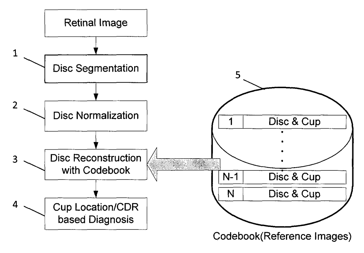
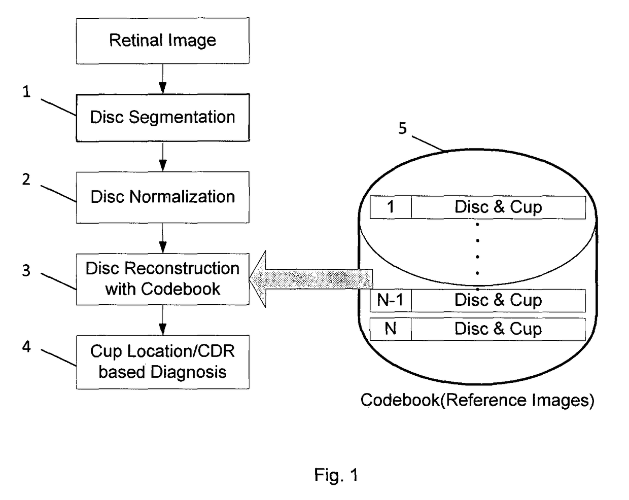
[0048]Referring to FIG. 1, the steps are shown of an embodiment of the invention, which performs Optic Cup Localization. As described in detail below the method employs cost-sensitive Linear Reconstruction.

[0049]The starting point of the embodiment is a single, non-stereo retinal image of an eye (typically a fundus image), referred to as the test image. In step 1, the optic disc of this eye is segmented within the test image using existing methods, for example Active Shape Model and template matching. Active shape matching is described at Yin, F., Liu, J., Ong, S. H., Sun, D., Wong, D. W. K., Tan, N. M., Baskaran, M., Cheung, C. Y., Aung, T., Wong, T. Y., Model-based Optic Nerve Head Segmentation on Retinal Fundus Images. In: IEEE Int. Conf. Engin. in Med. and Biol. Soc., pp. 2626-2629 (2011) . Template matching is described at, J., Liu, J., Wong, D. W. K., Yin, F., Cheung, C. Y., Baskaran, M., Aung, T., Wong, T. Y.: Automatic Optic Disc Segmentation with Peripapillary Atrophy Elimi...


PUM


Abstract

A method is presented to obtain, from a retinal image, data characterizing the optic cup, such as data indicating the location and / or size of the optic cup in relation to the optic disc. A disc region of the retinal image of an eye, is expressed as a weighted sum of a plurality of pre-existing “reference” retinal images in a library, with the weights being chosen to minimize a cost function. The data characterizing the cup of the eye is obtained from cup data associated with the pre-existing disc images and the corresponding weights. The cost function includes (i) a construction error term indicating a difference between the disc region of the retinal image and a weighted sum of the reference retinal images, and (ii) a cost term, which may be generated using a weighted sum over the reference retinal images of a difference between the reference retinal images and the disc region of the retinal image.

Description

CROSS-REFERENCE TO RELATED APPLICATIONS[0001]This patent application is a U.S. National Phase Application under 35 U.S.C. §371 of International Application No. PCT / SG2014 / 000026, filed Jan. 22, 2014, entitled COST-SENSITIVE LINEAR RECONSTRUCTION BASED OPTIC CUP LOCALIZATION, which claims priority to Singapore Patent Application No. 201300527-7, filed Jan. 22, 2013.FIELD OF THE INVENTION[0002]The present invention relates to methods and systems for processing at least one image of an eye, such as for measurement of a cup-to-disk-ratio (CDR). The CDR measurement may be used as part of a process for glaucoma diagnosis, optionally including also a step of treatment of glaucoma.BACKGROUND OF THE INVENTION[0003]Glaucoma is a chronic eye condition in which the nerve that connects the eye to the brain (optic nerve) is progressively damaged. Patients with early glaucoma do not have visual symptoms. Progression of the disease results in loss of peripheral vision, so patients may complain of “...

Claims


Application Information

Patent Timeline
Patent Type & Authority Patents(United States)
IPC IPC(8): G06T7/00G06T7/11G06K9/62G06K9/46G06K9/52
CPCG06T7/0014G06K9/46G06K9/52G06K9/6215G06T7/11G06K2009/4666G06T2207/30041G06F18/22
Inventor XU, YANWULIU, JIANGCHENG, JUNYIN, FENGSHOUWONG, WING KEE DAMON
Owner AGENCY FOR SCI TECH & RES
Why Eureka
Try Eureka