High intensity focused ultrasound device with a concave segment shaped transducer for eye treatment

Active Publication Date: 2016-12-13
EYE TECH CARE +1
View PDF47 Cites 0 Cited by
  • Summary
  • Abstract
  • Description
  • Claims
  • Application Information

AI Technical Summary

Benefits of technology

The patent describes a need for a safe and effective method of treating ocular pathologies using high intensity focused ultrasound (HIFU). The invention meets this need by providing a device that can easily and safely treat the whole circumference of the eye in only one step without the need for manipulation during the procedure. The device comprises an eye ring with a means to generate ultrasound beams on the distal end of the eye ring. The technical effect of this invention is to provide a precise, safe, and affordable treatment for ocular pathologies.

Problems solved by technology

In the field of ophthalmologic disease, it is well known that glaucoma is a significant public health problem, between 1 to 2% of population being suffering from this pathology, because glaucoma is a major cause of blindness.
When the first visual troubles appear, lesions are commonly already large and despite irreversible.
If the pressure remains high for a long period of time (many years), total vision loss occurs.
High pressure develops in an eye because of an internal fluid imbalance.
With the increased pressure in the eye, the aqueous fluid builds up because it cannot exit fast enough.
The increased IOP compresses the axons in the optic nerve and also may compromise the vascular supply to the optic nerve.
Currently available medications may have many serious side effects including: congestive heart failure, respiratory distress, hypertension, depression, renal stones, aplastic anemia, sexual dysfunction and death.
Compliance with medication is a major problem, with estimates that over half of glaucoma patients do not follow their correct dosing schedules.
In many cases, these operations are a temporary solution, as there is not yet a cure which is completely satisfactory for glaucoma.
Trabeculectomy is associated with many problems.
Failure from scarring may occur, particularly in children and young adults.
The use of these agents has increased the success rate of trabeculectomy but also has increased the prevalence of hypotony.
Hypotony is a problem that develops when aqueous flows out of the eye too fast.
Endophthalmitis often leads to permanent and profound visual loss.
The bleb can tear and lead to profound hypotony.
The bleb can be irritating and can disrupt the normal tear film, leading to blurred vision.
The major limitation of this technique is that it is a very difficult surgical technique and only a few surgeons are able to perform it successfully.
Many complications are associated with aqueous shunt devices.
The problem with cyclophotocoagulation is that one shot destroys only a small region of ciliary body and many shots are necessary all around the eye globe, so that a sufficient part of the ciliary body is destroyed.
This manual technique is quite empiric, non reproducible, long and not easy.
This method presents the inconvenient that it is necessary to repeat the operation (i.e. rotate the inner cylinder, acquire an image, verify that the apparatus is still in place, produce a laser shot) many times to treat the whole region to be treated.
Furthermore, this method may induce damages to the visual functions (due to spot size errors, misalignment between the ultrasonic head, the laser means and the fixing means, etc.).
Finally, the need of repeating an operation many times increases the operative time and thus the error risk factor.
However, such apparatus which was manufactured and distributed under the commercial name of SONOCARE was very difficult to manipulate.
Thus—as disclosed above with regard to laser techniques—each shot needs to be repeated many time to treat all the circumference of the eye and all the apparatus needs to be handled, placed and calibrated many times, thus taking a very long time (i.e. displacement of the ultrasonic means, verification of the position of the ultrasonic means with regard to the punctual region to be treated with optical and echographic sighting means, filling of the device with coupling liquid and production of a ultrasonic shot).
Even if this method provides a treatment to glaucoma in a non invasive manner, it presents the inconvenient that it is necessary to repeat the operation many times to treat the eye circumferentially.
Moreover, tissues at the neighbourhood of the treatment area can be destroyed leading to blurred vision, eye muscle imbalance or double vision.
It is consequently hard and expensive to apply this method in the treatment of glaucoma.

Method used


Image

  • High intensity focused ultrasound device with a concave segment shaped transducer for eye treatment
  • High intensity focused ultrasound device with a concave segment shaped transducer for eye treatment
  • High intensity focused ultrasound device with a concave segment shaped transducer for eye treatment

Examples


Embodiment Construction

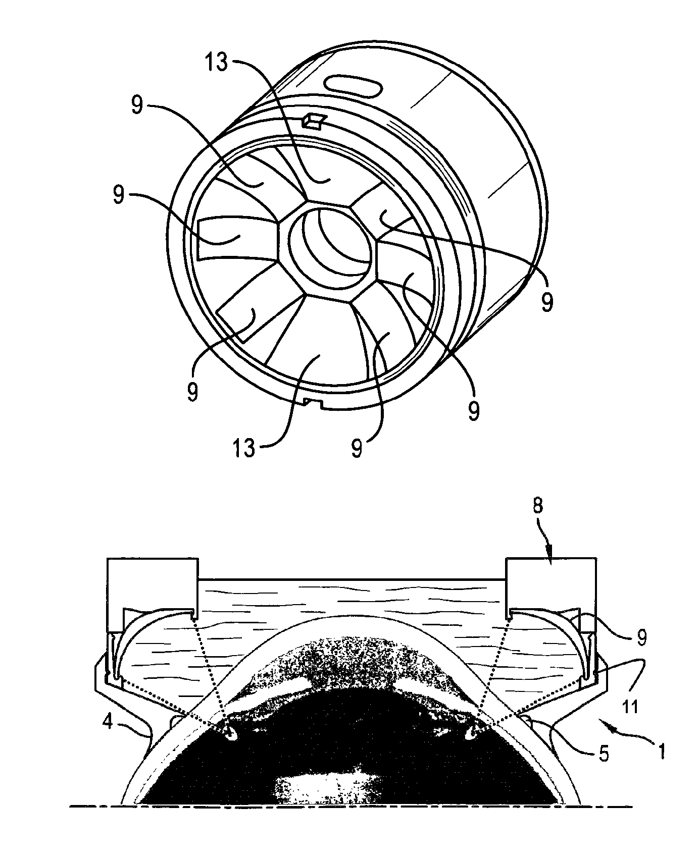
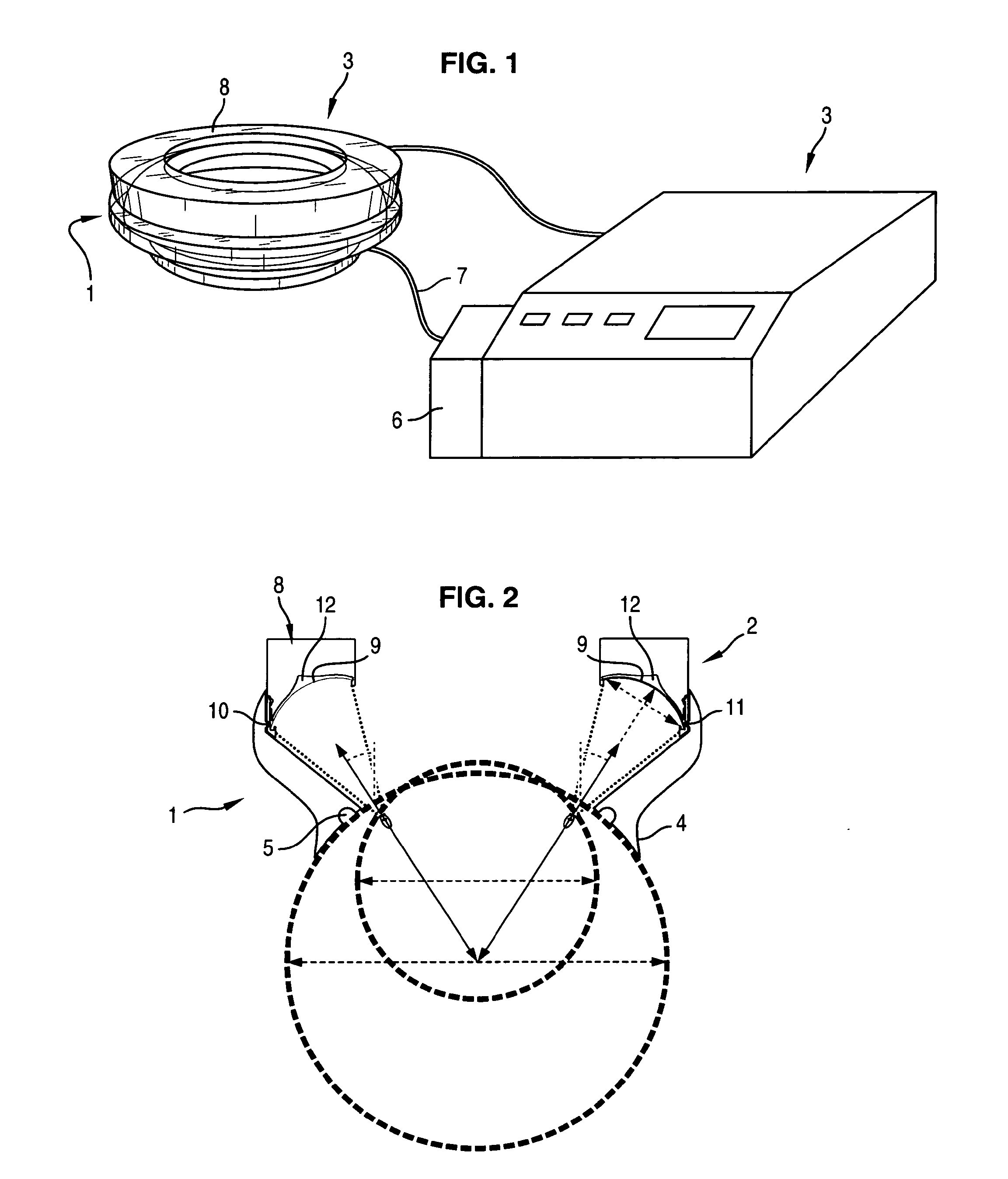
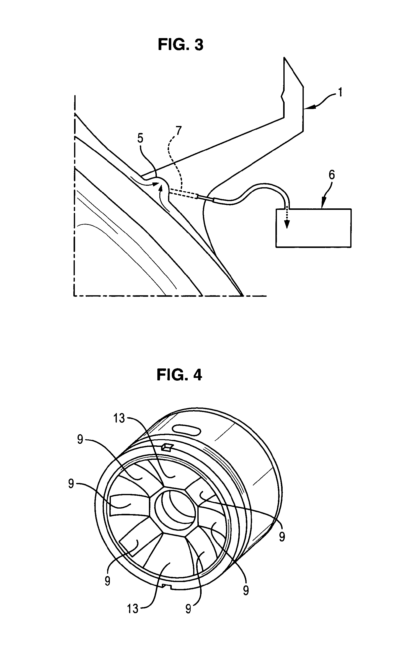
[0056]According to another embodiment of the invention, said means fixed on the distal end of the eye ring are suitable to generate scattered ultrasound beam.

[0057]The eye ring consists in a sawn-off cone element open at both ends wherein the small base is the proximal end and the large base is the distal end.

[0058]The proximal end of the sawn-off cone element comprises an external annular flange suitable to be applied onto the eye globe.

[0059]The proximal edge of the sawn-off cone element comprises an annular groove communicating with at least one hose formed in the sawn-off cone element and connected to a suction device.

[0060]The internal diameter of the proximal end of the sawn-off cone element is sensibly equal to the corneal diameter plus 2 to 6 mm, and more preferably equal to the sum of the corneal diameter with a value of 4 millimeters.

[0061]The internal diameter of the proximal end of the sawn-off cone element, depending on the patient corneal diameter, can be comprised bet...


PUM


Abstract

A device for treatment of an ocular pathology characterized in that it comprises at least one eye ring (1) wherein the proximal end of said eye ring (1) is suitable to be applied onto the globe and a generator (2,17) to generate ultrasound beam fixed on the distal end of the eye ring (1), the generator to generate ultrasound beam presenting a concave segment shape conformed along a single curvature corresponding to a single direction wherein the concavity is designed to be tuned towards the eyeglobe.

Description

CROSS REFERENCE TO RELATED APPLICATIONS[0001]This application is a §371 National Stage Application of PCT / EP2009 / 060678 filed Aug. 18, 2009, which claims priority to PCT / EP2009 / 051892 filed Feb. 18, 2009.BACKGROUND OF THE INVENTION[0002]Field of the Invention[0003]The present invention is generally directed to a non invasive treatment for ocular pathology, and relates more particularly to a device and method for generating high intensity focused ultrasound onto at least one annular segment of the ciliary body of an eye affected by glaucoma[0004]Description of Related Art[0005]In the field of ophthalmologic disease, it is well known that glaucoma is a significant public health problem, between 1 to 2% of population being suffering from this pathology, because glaucoma is a major cause of blindness.[0006]The World health organisation considers glaucoma as the second cause of blindness in the world, responsible of 15% of declared blindness occurrences, with an incidence of 2.4 millions...

Claims


Application Information

Patent Timeline
IPC IPC(8): A61B8/14A61F9/007A61N7/02A61N7/00A61B18/00
CPCA61F9/00745A61N7/02A61B2018/00291A61F9/00781A61N2007/0065A61N2007/0078A61N2007/027
Inventor ROMANO, FABRIZIOLAFON, CYRILCHAPELON, JEAN-YVESCHAVRIER, FRANCOISEBIRER, ALAINFARCY, LAURENTCHAPUIS, PHILIPPE
Owner EYE TECH CARE
Why Eureka
Try Eureka