
Endoscope image-acquisition unit and endoscope apparatus

an endoscope and image acquisition technology, applied in the field of endoscope image acquisition units and endoscope apparatuses, can solve the problems of reducing the pixel pitch and the problem of assembly errors of several micrometers, and achieve the effect of exceeding the tolerances

Active Publication Date: 2016-06-14
OLYMPUS CORP
View PDF32 Cites 0 Cited by
  • Summary
  • Abstract
  • Description
  • Claims
  • Application Information

AI Technical Summary

Benefits of technology

The patent text describes a way to make sure that an image-capturing device is in the correct position when using a microscope or other optical instruments. This is important because it helps to get better and more accurate results from the experiments or observations that are being done. The technique helps to overcome any mistakes that might occur during the manufacturing process of the optical instruments.

Problems solved by technology

Associated with this decrease in the pixel pitch, allowable assembly errors between individual lenses, between an image-acquisition device and an objective lens, and so forth are also decreasing, and thus, assembly errors of several micrometers are becoming problematic.

Method used


Image

  • Endoscope image-acquisition unit and endoscope apparatus
  • Endoscope image-acquisition unit and endoscope apparatus
  • Endoscope image-acquisition unit and endoscope apparatus

Examples


Embodiment Construction

[0018]An endoscope image-acquisition unit according to an embodiment of the present invention will be described below with reference to the drawings.

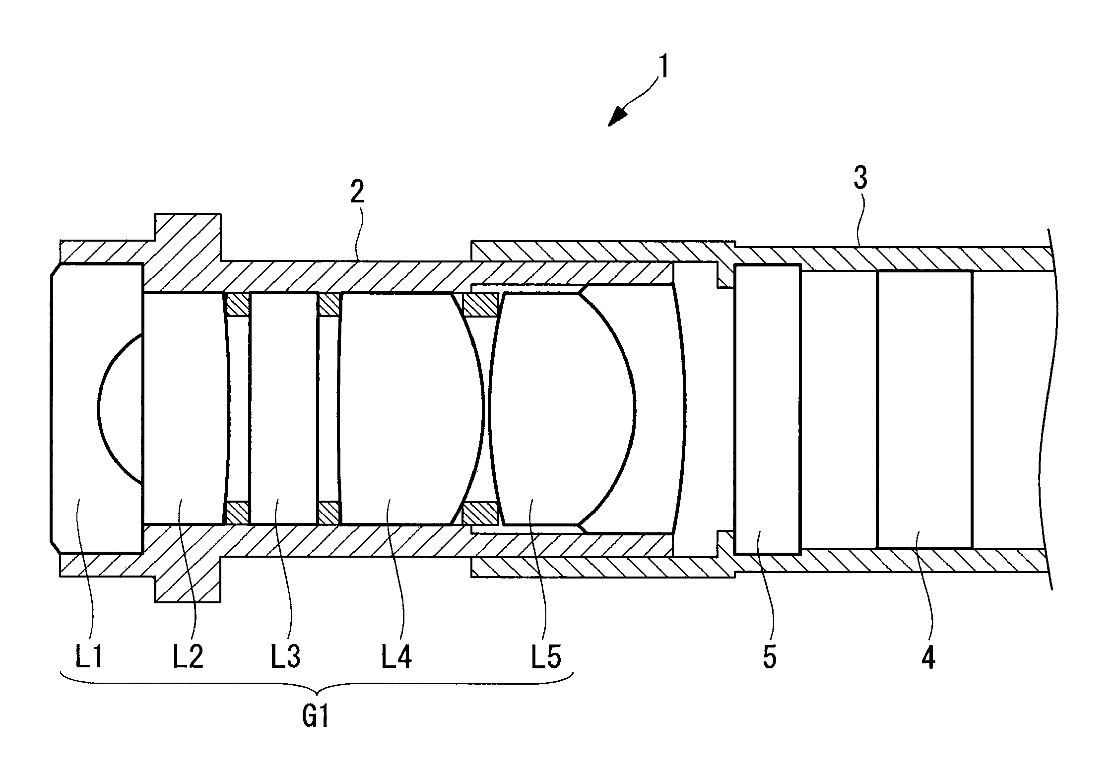
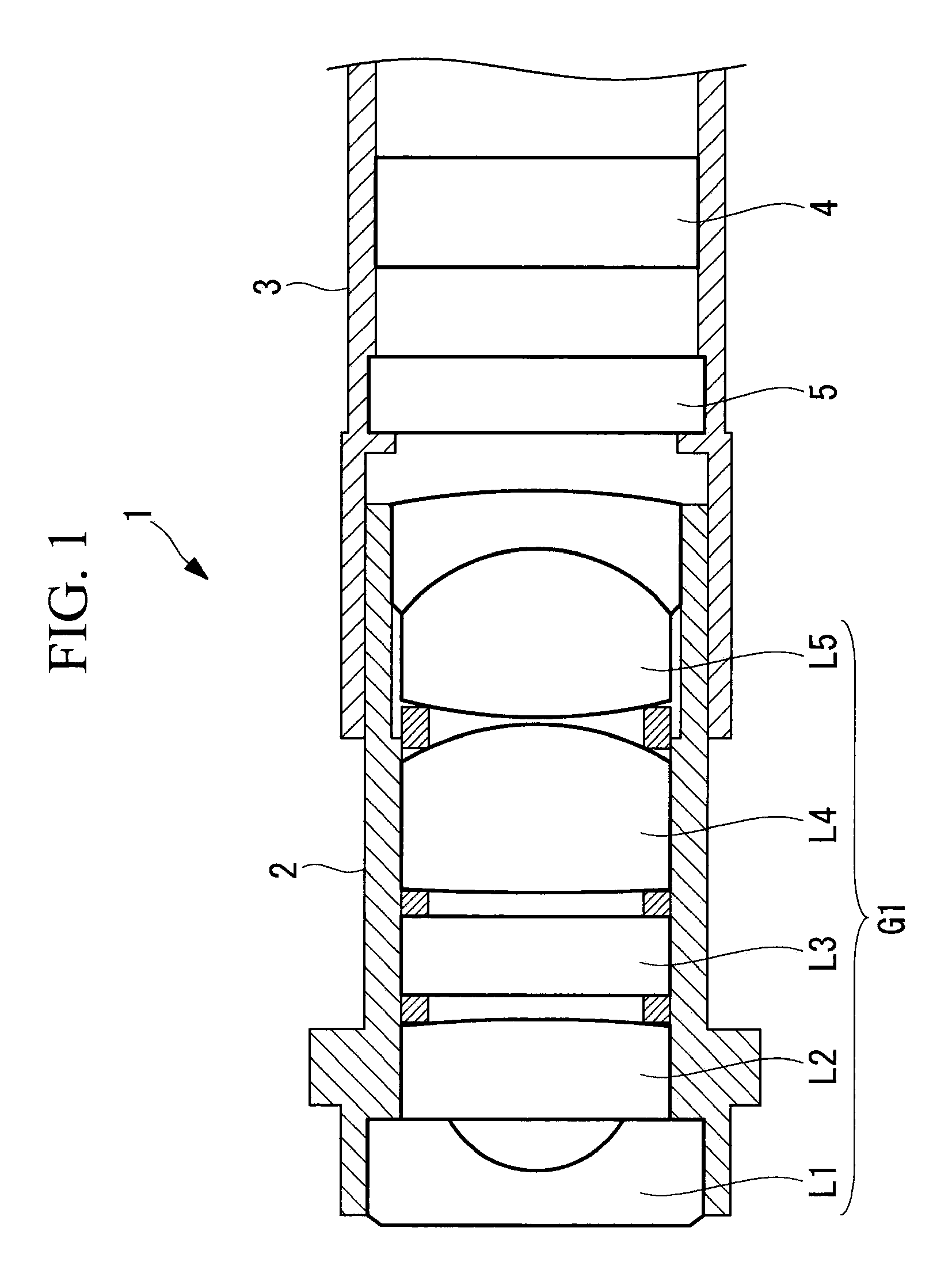
[0019]An endoscope image-acquisition unit 1 according to this embodiment shown in FIG. 1 is provided with an objective-lens-unit frame 2 that holds an objective lens and an image-acquisition-device holding frame 3 that is fitted to the objective-lens-unit frame 2 and that holds an image-acquisition device.

[0020]The objective-lens-unit frame 2 holds a lens group G1 formed of a plurality of objective lenses L1, L2, L3, L4, and L5.

[0021]The image-acquisition-device holding frame 3 holds an image-acquisition device 4 and a cover glass 5. In addition, the image-acquisition-device holding frame 3 is formed of polysulfone so as to allow microwaves to pass therethrough. Note that the material that forms the image-acquisition-device holding frame 3 is not limited to polysulfone so long as the material allows microwaves to pass therethrough. Ther...


PUM


Abstract

Positioning precision between an image-acquisition device and an objective lens is enhanced by suppressing manufacturing errors. Provided is an endoscope image-acquisition unit equipped with an objective-lens-unit frame that holds an objective lens and an image-acquisition-device holding frame that is fitted to the objective-lens-unit frame and that holds an image-acquisition device, wherein the objective-lens-unit frame and the image-acquisition-device holding frame are attached and secured to each other by means of thermosetting resin that is applied to fitting portions therebetween and in which polar-molecule materials are mixed, and one of the objective-lens-unit frame and the image-acquisition-device holding frame, whichever one is positioned outside at the fitting portions, is formed of a material that allows microwaves to pass therethrough.

Description

CROSS-REFERENCE TO RELATED APPLICATIONS[0001]This is a continuation of International Application PCT / JP2014 / 066365, with an international filing date of Jun. 19, 2014, which is hereby incorporated by reference herein in its entirety. This application claims the benefit of Japanese Patent Application No. 2013-130519, the content of which is incorporated herein by reference.TECHNICAL FIELD[0002]The present invention relates to an image-acquisition unit employed in an endoscope and to an endoscope apparatus provided with this image-acquisition unit.BACKGROUND ART[0003]In medical endoscopes such as nasal endoscopes or the like, there has been a demand in recent years for reducing the size of a tip of an inserted portion thereof from the viewpoint of reducing the burden on patients or the like. In response, small image-acquisition devices (CCDs, and CMOSs) have been developed for endoscopes, and the pixel pitch thereof is also decreasing year by year. Associated with this decrease in the...

Claims


Application Information

Patent Timeline
Patent Type & Authority Patents(United States)
IPC IPC(8): A61B1/00A61B1/04G02B7/02G02B23/24G01J1/02G01J1/04
CPCA61B1/04A61B1/0011A61B1/00105A61B1/00128A61B1/00195G01J1/0271G01J1/0411G02B7/02G02B7/025G02B23/243G02B23/2476A61B1/00096
Inventor SASAMOTO, TSUTOMUMURATA, KEIJI
Owner OLYMPUS CORP
Learn More