MiR-33 inhibitors and uses thereof to decrease inflammation
a technology of anti-inflammatory molecules and inhibitors, applied in the field of molecular biology, can solve problems such as tissue injury, and achieve the effect of increasing the stability of anti-sense oligonucleotides
- Summary
- Abstract
- Description
- Claims
- Application Information
AI Technical Summary
Benefits of technology
Problems solved by technology
Method used
Image
Examples
example 1
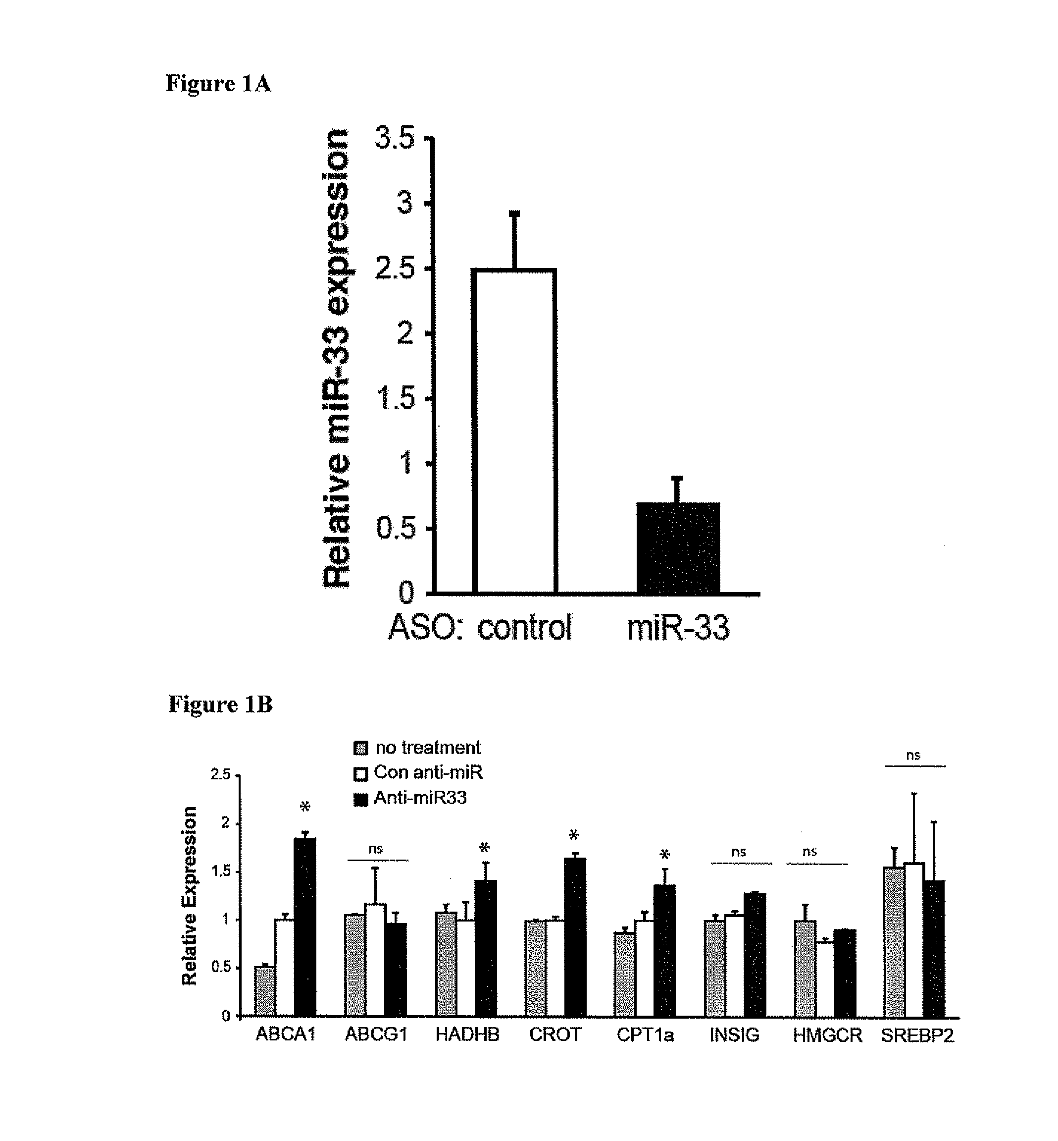
Anti-miR33 Increases Expression of miR-33 Target Genes in the Liver, including ABCA1
[0174]Materials and Methods
[0175]Mice
[0176]All experiments were approved by the New York University School of Medicine Institutional Animal Care and Use Committee. LdIr− / − mice were weaned at 4 weeks of age and placed on a high fat diet (21% (wt / wt) fat, 0.3% cholesterol; Research Diets) for 14 weeks, at which point mice were either sacrificed (baseline) or switched to chow diet for 4 weeks. Coincident with the switch to chow diet, mice were randomized into 3 groups (n=15 mice): no treatment (PBS), 2′F / MOE control anti-miR or 2′F / MOE anti-miR33 oligonucleotide (Regulus Therapeutics). Mice received 2 injections of 10 mg / kg anti-miR (or an equivalent volume of PBS) the first week, spaced two days apart, and weekly injections of 10 mg / kg anti-miR (or PBS) thereafter for 4 weeks. Following the second injection of anti-miR, mice were monitored for inflammation by measurement of serum IL-6 and MCP-1 by ELI...
example 2
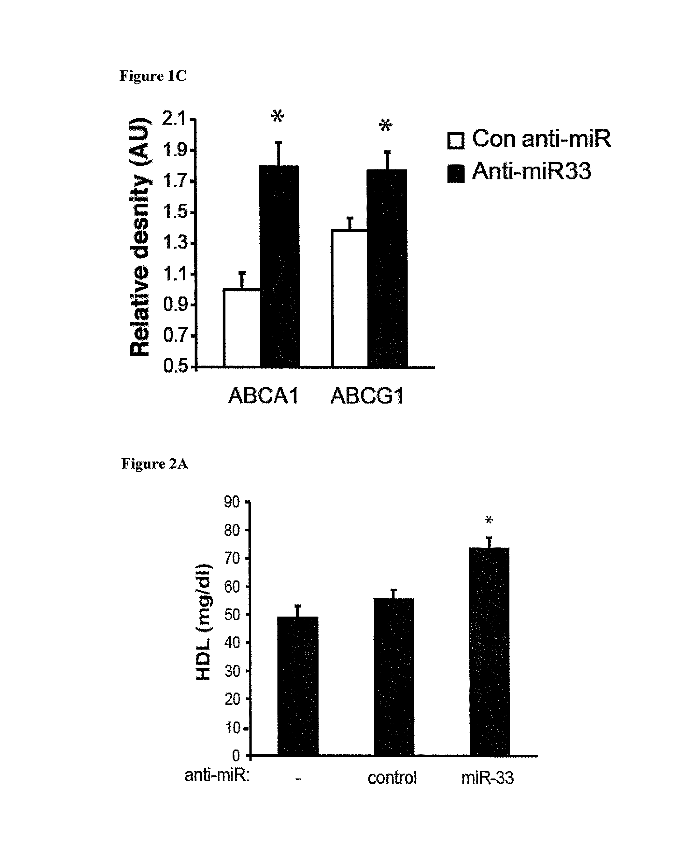
Anti-miR33 Treatment Increases HDL and Enhances Reverse Cholesterol Transport In vivo
[0184]Materials and Methods
[0185]Plasma Lipoprotein Analysis
[0186]Plasma was collected at sacrifice and total cholesterol was assayed (1:5 dilution) using the Cholesterol-E kit (Wako) as described (28). For FPLC analysis, 300 μl of pooled plasma (n=8 mice total) was separated on a Superose column (Amersham) at a flow rate of 0.4 ml / min as described (Rayner, K J., et al. 2010. Science 328:1570-1573). Fractions were collected and analyzed for total cholesterol content using the Cholesterol-E kit. For HDL measurements, apoB-containing lipoproteins were precipitated by the phosphotungstate-magnesium method, and HDL cholesterol was measured using either the HDL Cholesterol kit (Wako) or the Amplex Red Cholesterol Assay (Invitrogen) (Rayner, K. J., et al. 2010. Science 328:1570-1573).
[0187]In vivo Reverse Cholesterol Transport Assay
[0188]Bone marrow derived macrophages were prepared from C57Bl / 6 mice as p...
example 3
Anti-miR33 Treatment Induces Atherosclerosis Regression and Lesion Remodeling
[0193]Materials and Methods
[0194]Atherosclerosis Analysis
[0195]Hearts embedded in OCT were sectioned through the aortic root (8 μm), and stained with hematoxylin and eosin for lesion quantification or used for immunohistochemical analysis as previously described (Moore, K. J., et al. 2005. J Clin Invest 115:2192-2201; Manning-Tobin, J. J., et al. 2009. Arterioscler Thromb Vasc Biol 29:19-26). For morphometric analysis of lesions, 16 sections per mouse were imaged, spanning the entire aortic root, and lesions were quantified using iVision Software. For collagen analysis, 10 sections per mouse were stained with Picrosirius Red and imaged under polarized light using a Zeiss Axioplan microscope. For detection of neutral lipid, oil red O staining was performed as previously described (Moore, K. J., et al. 2005. J Clin Invest 115:2192-2201; Manning-Tobin, J. J., et al. 2009. Arterioseler Thromb Vasc Biol 29:19-26...
PUM
| Property | Measurement | Unit |
|---|---|---|
| pH | aaaaa | aaaaa |
| flow rate | aaaaa | aaaaa |
| stability | aaaaa | aaaaa |
Abstract
Description
Claims
Application Information
 - R&D
- Intellectual Property
- Life Sciences
- Materials
- Tech Scout
- Unparalleled Data Quality
- Higher Quality Content
- 60% Fewer Hallucinations
Browse by: Latest US Patents, China's latest patents, Technical Efficacy Thesaurus, Application Domain, Technology Topic, Popular Technical Reports.
© 2025 PatSnap. All rights reserved.Legal|Privacy policy|Modern Slavery Act Transparency Statement|Sitemap|About US| Contact US: help@patsnap.com


