Eureka AIR delivers breakthrough ideas for toughest innovation challenges, trusted by R&D personnel around the world.

Device for the removal of thrombi

a technology for thrombosis and thrombosis, which is applied in the field of thrombosis removal devices, can solve the problems of affecting the function of the pulmonary artery, the inability to supply oxygen and nutrients to the associated tissue, and the destruction of relevant tissue, etc., and achieves the effect of excellent attachment or “adhesion”

Active Publication Date: 2015-06-16
PHENOX
View PDF26 Cites 12 Cited by
  • Summary
  • Abstract
  • Description
  • Claims
  • Application Information

AI Technical Summary

Benefits of technology

[0013]The invention is based on findings by the inventors proving that the use of such a simply designed device capable of being made of particularly small size is suited to recover thrombi also from vessels having an especially small lumen. The guide wire designed as insertion aid enables the device to be easily moved also into small-lumen and convoluted vessel segments and keeps the overall diameter of it small, but in particular the device diameter that cannot be varied (the diameter which is brought about by the fibers may be variable in as much as these are flexible so that even constricted vessel routes can be passed through). The fibers / bristles are suited to capture and stabilize a thrombus, especially if they are made of or finished with thrombogeneous materials.
[0016]It is understood that for the intended purpose the fibers / bristles must have adequate stiffness but at the same time must be flexible or bendable enough so that they can be passed through a catheter and do not damage the vessel walls.
[0027]In accordance with another expedient embodiment the fibers have a hook-shaped configuration with their hook-shaped ends advantageously being bent in proximal direction so that when being inserted into the blood vessel they cannot impede the insertion process but serve to improve thrombus anchoring during retraction from the blood vessel. Furthermore, crimpy or helix-shaped structures may be employed as well. As far as the fibers are intended and suited to be “glued” to a thrombus good clinging properties and a large contact area are considered to be beneficial.
[0031]Especially preferred is an angle of 90° maximum or slightly less than 90° between the distal element and the longitudinal axis of the fibers. The embodiment providing for an angle of slightly less than 90° is particularly atraumatic when moving into the vessel or through the tromb and at the same time results in an especially effective anchoring within the thrombus when the element is pulled out of the blood vessel.
[0042]As per a particularly preferred embodiment of the device the fibers are coated. For example, this coating may be a neutral one consisting of Parylene or Teflon®, but may also comprise collagen or may be a coating of a material conducive to blood coagulation, preferably having one or several coagulation factors. This embodiment serves to strengthen the anchorage of the fibers inside the thrombus and alleviates the risk of the thrombus disintegrating leaving fragments of the thrombus in the blood vessel or allowing them to be released in the blood stream.
[0055]As per another beneficial embodiment the braces of the cage structure are arranged in a helical fashion, i.e. starting point and end point on the distal element are offset against each other by a certain angle, ranging for example between 45° and 180°, preferably by approx. 90°. Such a helical line arrangement allows a thrombus adhering to a vessel wall to be sheared or cut off when the cage structure is moved forward without the necessity of having to turn to device.

Problems solved by technology

The obstruction of organ arteries also leads to supply of oxygen and nutrients to the associated tissue being interrupted.
The disorder of the functional metabolism linked with functional losses is closely followed by a failure of the structural metabolism resulting in the relevant tissue becoming destroyed (infarction).
Nevertheless, the arteries of the limbs as well as pulmonary arteries are also impaired.
The disease pattern of the thrombotic occlusion of an intracranial sinus may lead to severe intracerebral hemorrhage due to a failure of venous drainage of brain tissue.
Since treatment methods of this kind are usually very time consuming they are frequently combined with invasions aimed at reducing the size of or removing the thrombus or embolus mechanically.
Disadvantages associated with the known transluminal devices are that with said devices it is often impossible to remove the thromb completely and, moreover, there is a risk of the thromb or fragments of it being released into the blood stream thus passing on to vessels of smaller lumen which are more difficult to reach and treated.
Furthermore, due to their size and / or low flexibility the devices known from prior art are only inadequately suited for the removal of thrombi from greatly convoluted vessels or those of particularly small lumen such as those in the brain.
The drawback associated with this device is, however, that in order to cool and thus keep the capture arms below transformation temperature before they are released into the blood stream it must either be moved behind the thrombus in a secondary catheter which brings about the cooling effect or inside the catheter provided with the capture arms a heating system has to be arranged that enables the transformation temperature to be attained when the thrombus has been reached.
Not only are the design requirements of this configuration very high and thus prone to disturbances it is also the sheer physical size of this device that rules out a treatment of vessels having a particularly small lumen.

Method used


Image

  • Device for the removal of thrombi
  • Device for the removal of thrombi
  • Device for the removal of thrombi

Examples


Embodiment Construction

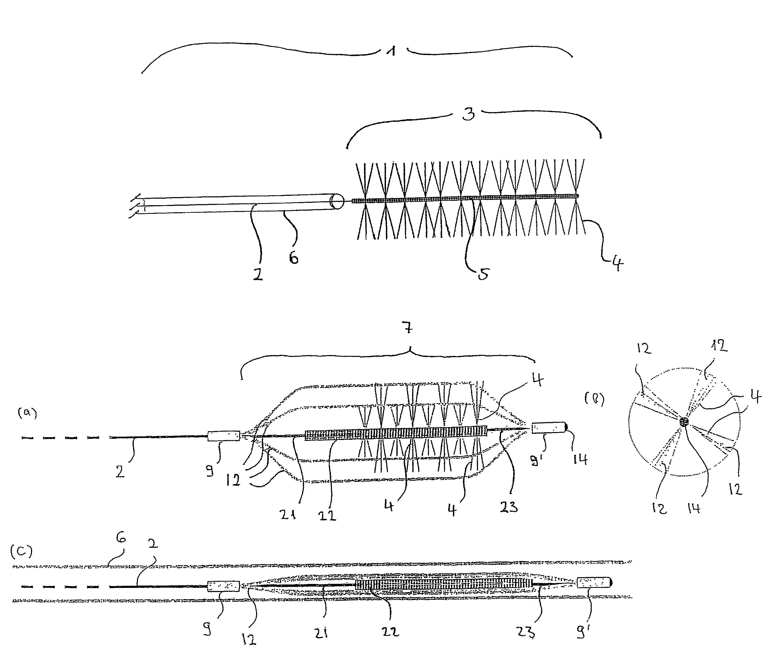
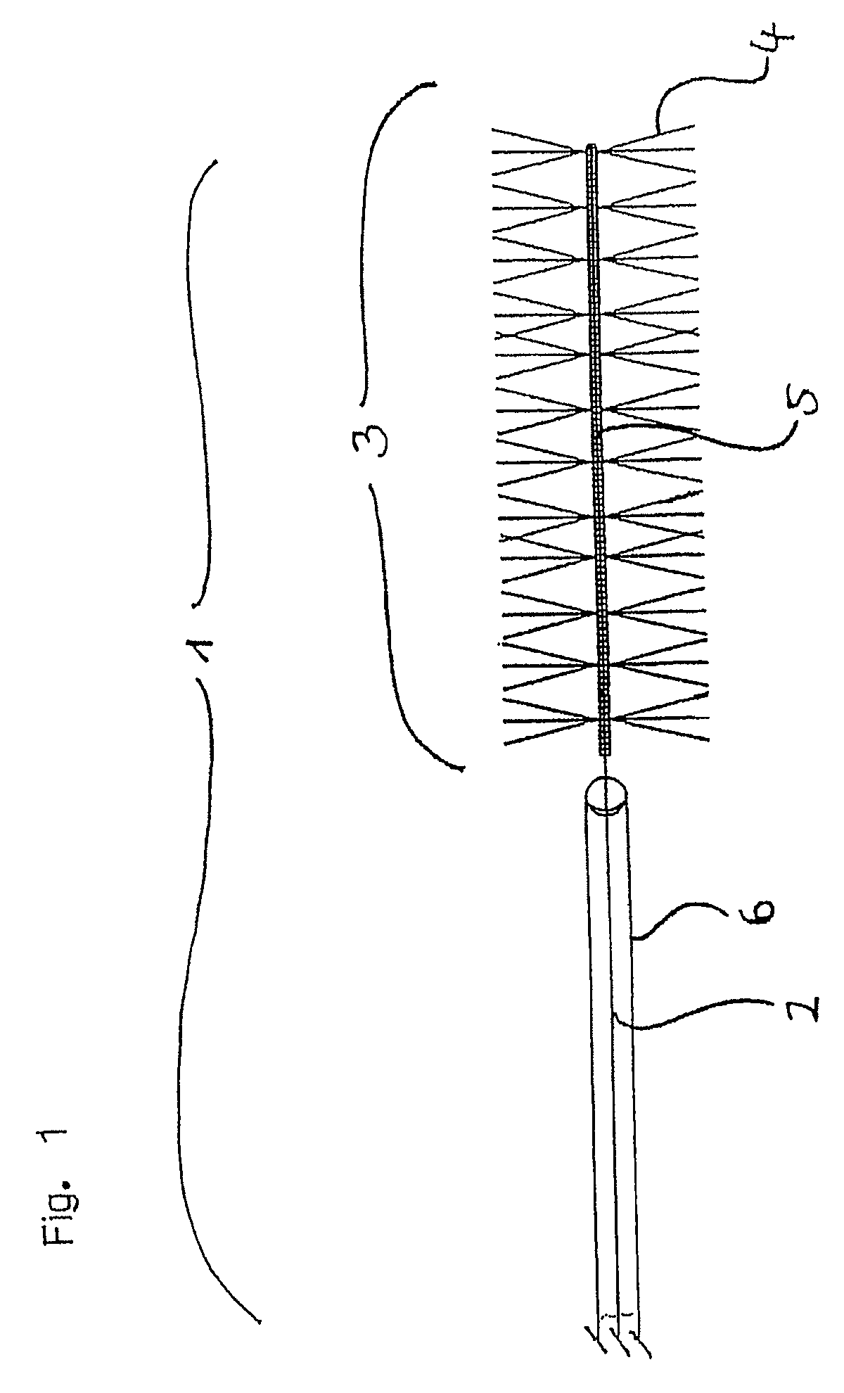
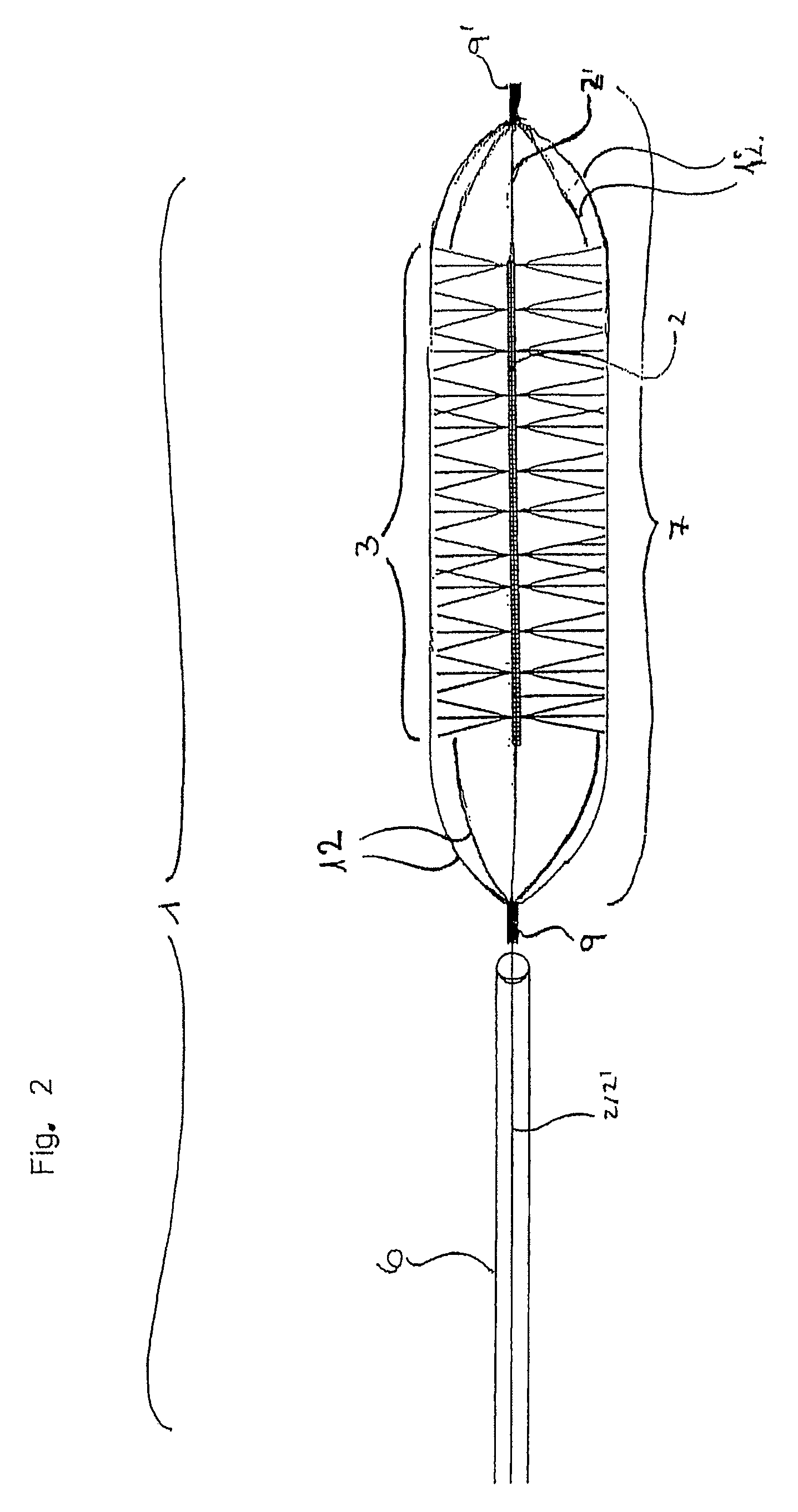
[0072]The inventive device 1 shown in FIG. 1 is provided at the distal end of the guide wire 2 made of nitinol and having a diameter of 0.254 mm with functional unit 3 intended for the retrieval of thrombi. Functional unit 3 is provided with polyamide bristles 4 having a length of 2 mm and being attached radially onto guide wire 2. The diameter of functional unit 3 is thus 4 mm and is particularly suited for the retrieval of thrombi out of vessels having an inner diameter ranging approx. between 4.5 and 5 mm. In the area where the bristles are situated guide wire 2 is designed as a platinum micro-coil 5; this embodiment is especially flexible and at the same time serves as radiopaque marker to enable the placement to be performed under radiographic observance. The distal tip of the micro-coil has been rounded off to render it particularly atraumatic. Bristles 4 are mechanically clamped into the coil with one fiber each forming two bristles 4, having a length slightly more than doubl...


PUM


Abstract

The invention relates to a device for the removal of foreign bodies and thrombi from body cavities and blood vessels using a guide wire provided with a distal element with said distal element being provided with an orthogonal structure.

Description

CROSS-REFERENCE TO RELATED APPLICATIONS[0001]This application is a continuation of PCT / EP2005 / 009057, filed Aug. 22, 2005, which claims priority from Germany Application No. 10 2004 040 868.8, filed Aug. 23, 2004, all of which are hereby incorporated herein by reference.BACKGROUND OF THE INVENTION[0002]The invention relates to a device for the removal of foreign bodies and thrombi from body cavities and blood vessels using a guide wire provided with a distal element. Said distal element is non-detachably connected with the guide wire.[0003]The invention, furthermore, relates to a combination comprising such a device and a cage element.[0004]Thromboembolic diseases such as cardiac infarction, pulmonary embolism, peripheral thrombosis, organ embolisms etc. are typically caused by a thromboembolism (hereinafter for short thromb or thrombus), i.e. a visco-elastic blood clot comprising platelets, fibrinogen, coagulation factors etc. forming in a blood vessel which it obstructs either who...

Claims


Application Information

Patent Timeline
Patent Type & Authority Patents(United States)
IPC IPC(8): A61B17/22A61B17/221A61B10/02A61B19/00
CPCA61B17/221A61B2010/0216A61B2017/22034A61B2017/22038A61B2017/2212A61B2019/547A61B2090/397A61B2017/22001A61B2017/22042A61B2017/22079
Inventor MILOSLAVSKI, ELINAHENKES, HANSHANNES, RALFMONSTADT, HERMANN
Owner PHENOX
Why Eureka
Eureka Blog
Learn More